Q. A patient presented to my office with optic neuritis. Should I comanage her with a neurologist to rule out MS?

A. There is a well-established link between optic neuritis and multiple sclerosis (MS). Although the exact risk of developing MS after a bout of optic neuritis is unknown, studies have reported anywhere from a 13% increased risk to an 88% risk, says Laura Falco, O.D., of Nova Southeastern University College of Optometry in Fort Lauderdale, Fla.
The Optic Neuritis Treatment Trial (ONTT) showed that 30% of all patients with optic neuritis develop MS within five years.1 Acute, idiopathic optic neuritis may represent a demyelinating event of the optic nerve. This may be the first clinical sign of MS, explains Leonard Messner, O.D., vice president for patient care at Illinois College of Optometry. Also, be suspicious if your patient has recurrent optic neuritis, as this is more common in MS patients.1
Patricia Modica, O.D. of State University of New York College of Optometry, says, The typical optic neuritis patient is young and healthy and presents with painful vision loss.
Expedient diagnosis is essential since research has shown early treatment may delay the onset of MS. At three years follow-up, the Controlled High-Risk Avonex Multiple Sclerosis Prevention Study (CHAMPS) documented a 66% risk reduction for developing MS among patients who were treated within the first month following an optic neuritis attack.2
Refer the patient for magnetic resonance imaging (MRI) to rule out silent white matter changes in the central nervous system. These indicate disseminated demyelination consistent with MS. White matter changes are the single most important risk factor in predicting which patients are at greatest risk of developing MS, says Dr. Modica.
Indeed, 10-year follow-up data from the ONTT indicated optic neuritis patients who demonstrate one or more white matter lesions on MRI have a 56% chance of developing clinically definite MS. Patients with no evidence of white matter disease had a 22% risk of developing MS in the same period.1
Some tests you can perform may also aid in the diagnosis, Dr. Falco says. Cranial nerve testing can yield important additional information, as can ocular motility, color vision testing and fundus examination.
Patients who present with atypical optic neuritis should undergo additional testing. It is important to identify this group of patients, because while the risk of MS is low, the risk of an underlying systemic disease is high, Dr. Modica says.
Atypical presentations include older patients, optic neuritis that is bilateral or symmetrical, accompanied by disc hemorrhage, vitritis, retinitis, uveitis or retinal exudation. Differential diagnosis should include collagen-vascular disease, sarcoidosis, syphilis, Lyme disease, tuberculosis and viral or bacterial infectious disease. If a laboratory work-up and chest X-ray are unproductive, lumbar puncture with cerebro-spinal fluid analysis may be indicated, she says.
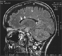
An MRI is a critical tool in diagnosising mutiple sclerosis.
This step requires comanagement with a neurologist. How-ever, because early treatment can limit the severity of MS, you should refer any suspect cases of optic neuritis for evaluation.
Q. What new systemic drugs are available for treating MS, and how well do they work?
A. Most patients with MS suffer from relapsing-remitting MS (RRMS) or secondary progressive MS (SPMS). RRMS patients have episodic attacks of MS about once a year and then go into a period of remission. SPMS patients have a more continuous degradation of neurological function. Over time, many RRMS sufferers report a shorter latency between demyelinating attacks associated with increased neurologic disability. Eventually some of these individuals show a steady deterioration of neurologic function and convert to SPMS, Dr. Messner says.
Interestingly, even when RRMS patients are is a period of remission, the inflammatory process in still active in the central nervous system, Dr. Falco adds. Therefore, clinical relapses do not accurately indicate the severity of the disease.
Independent of clinical symptoms, MS is a steadily progressive neurologic disorder that responds best to early anti-inflammatory and immunomodulatory treatment, Dr. Messner says.
The ONTT demonstrated that intravenous methylprednisolone improves the visual recovery time of optic neuritis in the short term, but has no beneficial effects on long-term visual recovery.1
In the last decade, a new era of therapy has provided more treatment options for patients with RRMS and SPMS. Interferon-beta drugs have immune-modulating properties that have proven useful in reducing relapses, delaying progression of disability and decreasing evidence of MS on MRI. Current studies believe that interferon reduces brain inflammation and inhibits new areas of inflammation, says Dr. Falco.
This class of drugs includes interferon Avonex (beta 1-a, Biogen), Betaseron (interferon beta 1-b, Berlex) and Copaxone (glatiramer acetate, Teva Pharmaceuti-cals). The CHAMPS study found that patients with optic neuritis and MRI evidence of white matter changes who were treated with Avonex were 44% less likely to convert to clinically significant MS. Dr. Modica notes that these patients also demonstrated a reduction in the volume of brainstem lesions and a reduction in new or enlarging lesions.2
Be aware that interferon therapy can result in adverse reactions, Dr. Falco says. Flu-like illness is the most common reaction, but it is reported to abate within 6-12 days of starting therapy.
Research is currently investigating other agents for treatment of MS as well. Antegren (natalizumab, Elan Pharmaceuticals), a humanized, monoclonal anti-adhesion molecule antibody, is currently in Phase III clinical trials for treatment of RRMS.3,4 This drug inhibits movement of immune cells from the bloodstream into the brain. Antegren is schedule to be filed for U.S. FDA approval by mid-2004.
1. Optic Neuritis Study Group. The 5-year risk of MS after optic neuritis. Experience of the Optic Neuritis Treatment Trial. Neurology 1997:49:1404-13.
2. OConner P, Controlled High-Risk Avonex Multiple Sclerosis Prevention Study (CHAMPS). The effects of intramuscular interferon beta 1-a in patients at high risk for the development of multiple sclerosis: a post hoc analysis of data from CHAMPS. Clin Ther 2003;25:2865-74.
3. Miller DH, Khan OA, Sheremata WA, et al. A controlled trial of natlizimab for relapsing multiple sclerosis. N Engl J Med 2003;348:15-23.
4. Dalton CM, Miszkiel KA, Barker GJ, et al. Effects of natalizumab on conversion of gaolinium enhancing lesions to T1 hypointense lesions in relapsing multiple sclerosis. J Neurol. 2004;251:407-13.
