 |
Sometimes, a condition that warrants our attention may also distract us from our obligation to consider the patients’ total ocular and visual status. Often, the identification of an ocular disease stops the diagnostic process short of fully identifying all of the patient’s needs, such as low vision management, prism or vision therapy. The presentation of deficiencies in visual acuity can sometimes be attributed to the effects of ocular disease as well as factors that fall within the scope of routine optometric practice. As such, optometrists must complete a thorough optometric evaluation notwithstanding the presence of ocular disease. This is especially important in the context of a case where the ocular disease limits visual acuity.
Surgical Shortcomings
A seven-year-old male was referred to us with a presentation of longstanding bilateral impairment of visual acuity, lateral movement of the head to the left side and increased frequency of squinting and closing the right eye. The patient had a history of esotropia and nystagmus at one year of age, which was addressed surgically in 2008 and was said to have resolved, subsequent to patching therapy that was ultimately unsuccessful. He was deficient in balance and depth perception, which was amplified in dark conditions. The patient’s medical and developmental histories were overall unremarkable.
 | |
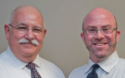
| The patient demonstrating the head turn found upon first presentation. He had to be told which direction his head used to turn as he did not remember. |
The patient’s mother observed that he had trouble walking up and down stairs, and on occasion preferred to crawl down them. He had difficulty distinguishing the borders of objects from a distance. She also indicated that he was having trouble with reading. Although the patient said that he did not have trouble seeing written material in his textbooks, his mother noted that he held reading material very close to his face. He also needed to sit in the front of the classroom and often walked up to the board in order to see the text. His mother observed that his movements appeared uncoordinated. He was in standard education classes and was underperforming, she said. His school was providing materials in large print, but was not being forthcoming with other compensatory strategies.
His entering corrected visual acuity was 20/80 at distance (OU). At near with both eyes together he saw 20/40 with a hyperopic astigmatic correction and flat-top bifocal (OD +4.25-3.00 x165, OD +4.00-4.00 x 180 OS, +2.75 add OU).
All chair skills were within normal limits except for the presence of nystagmus, which was seen when we performed eye movement testing was during which he showed significant fixation loss. Gross motor eye movement testing showed poor pursuit saccades. No improvement was noted in visual acuity with trial frame refraction. Phoroptor testing, cover test and NPC were not attempted secondary to the presence of nystagmus, although there was no gross deviation present by observation. The nystagmus was found to oscillate least when the patient had his head turned to the left. Anterior segment and indicated posterior segment evaluations were within normal limits.
Based on the previous primary care and current vision therapy examinations, the patient was diagnosed with decreased vision secondary to congenital nystagmus and oculomotor dysfunction. A three-pronged approach to treat the patient was put into effect.
1. Low Vision Treatment
The visual acuity loss was managed with traditional low vision devices.
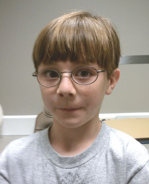 | |
| The patient with glasses, showing the lack of head turn. |
For distance, Max TV glasses (Eschenbach)—a 2x magnification system—were worn during school and for other distance activities. In office, visual acuity with the Max TV glasses was measured at 20/40 OU. The downside to using these glasses is that the patient had to remove his habitual prescription, but the acuity gain made this a moot point. When these were placed on the child’s face, he smiled and said that he had never seen so well.
For near vision, a 4x dome magnifier was used for all activities. In office, visual acuity with the dome magnifier was measured at 20/15 OU. This portable device allowed him comfort knowing that it would always be with him and could fit into his backpack or pocket. Additionally, a 2x bar magnifier with light was used for near activities involving reading, so that the patient can see the entire line at once. Patients with low vision often benefit from increased lighting. In this case, the contrast on the copied work was extremely poor, so the patient was excited.
2. Yoked Prism
A 4∆ yoked prism (prism with the bases in the same direction and of equal power) based left, was used to reduce the amount of head turn, essentially driving the patient into the null point.1,2 Yoked prism shifts the image in the same direction in each eye without impacting the image quality. For this child, reducing the amount of time that he was turning his head was crucial to avoiding issues with posture and neck/spinal issues in the future. The amount was determined by trial and error and, once determined, was trialed for two weeks using Fresnel prism. While the impact was immediately witnessed in the office, adaptation was a concern. Upon evaluation several weeks later, his mother indicated that the turn was still significantly reduced so the decision was made to grind in the prism.
 | |
| 4x dome and 2x bar magnifiers aided near vision tasks. |
3. Oculomotor-based Vision Therapy
Therapy was implemented to improve eye movements and the coordination of the visual motor system. Since the child was performing poorly on visually dependent activities, the therapy program encouraged better quality and more efficient visual input. Reading tutoring was a future consideration but vision therapy would set the stage for future success. Since many of the complaints were visual-motor based, the therapy program stressed gross motor visual skills at first, before moving to fine motor and reading eye movements. The therapy moved from larger to smaller targets and moved in sequence from monocular, bi-ocular to binocular skills. Fixation skills were addressed, then pursuits and finally saccades. Activities included but were not limited to Wayne Saccadic Fixator (an electronic board with lights that appear in set patterns and provides tactile and auditory feedback, Wayne Engineering), pegboard (stationary and rotating), chalkboard racetrack and stick in straw, starting with marker in paper towel roll.
This child’s visual acuity and efficiency issues, along with potential neck pain, might have been overlooked or disregarded. This case shows that blending several optometric disciplines allows practitioners to use all of the tools at their disposal to treat the visual and systemic signs and symptoms intrinsic to each of our patients.
1. Cotter SA. Clinical Uses of Prism: A Spectrum of Applications. St. Louis: Mosby, 289.2. Fischer V, Mahaphon T. Use of Yoked Prism in the Management of Nystagmus in Spina Bifida. Optometry 2006;77(6):275-6.
