We saw many examples of practitioners capitalizing on existing technology at this years Association for Research in Vision and Ophthalmology (ARVO) meeting. This trend was most evident in wavefront diagnostics, which is proving useful in cataract assessment, presbyopia, IOLs and contact lens wear. For those of you who comanage many patients, there are assessments of new surgical options for refractive complications and for patients who would traditionally require penetrating keratoplasty.
Heres a sampling of abstracts aimed at sharpening existing technologies and techniques.
Advances in Wavefront Diagnostics
Researchers continue to find new and practical ways to implement wavefront technology, especially to fine-tune vision correction and assessment.
Wavefront sensing provides a quick, non-invasive method of quantifying aberration associated with early-stage cataract formation, according to researchers at the University of Houston.1747 They used the Shack-Hartmann wavefront device to assess 149 patients. They measured for aberration, nuclear opalescence and various scatter metrics. Patients who had significant cortical and posterior subcapsular cataract were excluded. Patients underwent visual acuity testing with both high- and low-contrast letters in both high and low luminance, and researchers evaluated wavefronts ability to predict variance in each test. The study concluded that backscatter, forward scatter and wave aberration accounted for more than 50% of the variance found in low-luminance, high-contrast visual acuity testing.

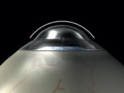
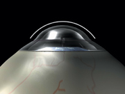
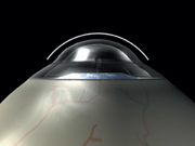
Conductive keratoplasty may be an option for patients who have had complications from other refractive surgery procedures.
Age-related changes in the lens affect not only accommodative amplitude but other aberrations as well, especially spherical aberration, according to a European study.1075 Researchers split 40 subjects into four ages groups: ages 20 to 30, 30 to 40, 40 to 50 and 50 to 60. Subjects underwent wavefront measurements in an unaccommodated state and in accommodations from 0 to 5.00D. Spherical aberrations varied significantly with age. The average spherical aberration coefficient decreased with increasing accommodation at a young age, reaching 0 for a stimulation of 3.00D. The same aberration was reversed in the older group, increasing under heightened accommodations.
Wavefront technology can also be used to assess patients who complain of triplopia (triple vision) associated with cataracts, a Japanese study finds.1748 Researchers at Osaka University Hospital used the Hartmann-Shack device to examine eight eyes of six patients who complained of monocular triplopia. All eyes showed mild nuclear cataract with myopia ranging from -7.50D to -19.00D. Wavefront analysis showed that the triple configuration was caused by an increase of trefoil and spherical aberration caused by the cataract.
Intraocular lenses with a prolate anterior surface, which was designed according to wavefront measurements proved beneficial in two studies.1751, 1752
In one study, researchers at Stanford and the Smith-Kettlewell Eye Research Center concluded that prolate intraocular lenses improve postoperative contrast sensitivity in cataract patients when compared to conventional IOLs.1751 Twenty-one patients (32 eyes) underwent cataract surgery. Control subjects received standard spherical IOLs, and the remaining patients received anterior-modified prolate IOLs. A Wilcoxon signed-ranks test for matched pairs showed a statistically significant improvement in contrast sensitivity scores for the patients with prolate intraocular lens implants.
Meanwhile, a study team at Loma Linda University found that prolate IOLs are better at correcting spherical aberrations than conventional IOLs.1752 Researchers compared 23 eyes implanted with a prolate lens with 23 eyes implanted with a conventional one. Although the refractive accuracy was higher with the conventional lens and these eyes displayed better uncorrected visual acuity, the eyes implanted with the prolate lens showed decreases in fourth-order spherical aberration. Theoretically, this should produce better contrast sensitivity. Additionally, the prolate lens appeared to decrease other higher-order aberrations, including horizontal coma and fourth-order astigmatism when compared with the conventional lens.

Patients with endothelial dystrophies may avoid spherical and corneal changes by undergoing deep lamellar endothelial keratoplasty.
Contact lens wearers can also benefit from wavefront assessment, concludes a University of Rochester study.1078 Based on wavefront aberration measurements, six patientsthree with normal corneas and three with abnormal corneas (one post-LASIK and two keratoconic)were fitted for custom contact lenses. When they compared normal with customized corrections, the researchers found that normal eyes with customized corrections improved an average of one half a line in high-contrast visual acuity and one line in low-contrast visual acuity. Abnormal eyes improved two lines in high-contrast visual acuity and three lines in low-contrast visual acuity.
Refractive Research
Are your presbyopic patients asking about laser surgery? An Israeli study found that LASIK-induced monovision was well tolerated by presbyopic patients, except for a small group who found night driving more difficult.2818 Investigators looked at 114 patients who had monovision post-LASIK, and discovered that 77% had binocular visual acuity of 20/25 or better for distance, and 87% had binocular visual acuity of J2 or better for near. Median stereo acuity was 100 seconds of arc (range 40 to 800 seconds of arc). Responses to questionnaires indicated that mean patient satisfaction was 80%. Also, 90% of patients considered their main goal met, but 15% reported increased difficulty driving at night.
Conductive keratoplasty (CK) might be a beneficial option for patients who had refractive surgery complications in whom flap manipulation or laser re-treatment is contraindicated, according to a study conducted by the Cornea & Laser Eye Institute in Teaneck, N.J.184 Six patients who had LASIK or PRK complications underwent CK and were evaluated for visual acuity, keratometry, topographic analysis and subjective assessment of visual quality. All showed improved visual acuity and improved quality of the corneal surface. They also reported reduced glare and halo symptoms.
As part of its evaluation of whether to provide refractive surgery to its aviators, the U.S. Army sponsored a study comparing LASIK and PRK under night vision goggle (NVG) conditions.2824 Twenty pilots underwent testing preoperatively, and at one week, one month and six months after randomized PRK or LASIK. Researchers measured high-contrast acuity and contrast sensitivity at 1, 2, 4, 8 and 16c/deg under quarter-moon and overcast starlight conditions. Under quarter-moon conditions, PRK subjects showed a decrease in NVG visual acuity at one week compared with LASIK, probably due to the epithelial healing process. After six months of healing, performance for both groups was equal in all conditions except extremely low starlight, in which mid-frequency contrast sensitivity in LASIK subjects was lower.
Implantable contact lenses (ICLs) produce a greater effect on the size of the retinal image than researchers anticipated, a study at the Vienna Medical University found.2817 Investigators looked at 65 eyes of 37 patients who received ICLs. Based on various ocular measurements, researchers estimated the pre- and postoperative retinal image sizes of distance objects and compared the image sizes to the change in clinical visual acuity. Calculations indicated that retinal image size should change (increasing for myopia, decreasing for hyperopia) 1.5% for every diopter of ametropia corrected. However, post-operative changes in visual acuity were higher than the researchers calculated: 7.1% higher for the 62 myopic eyes and 9.5% higher for the three hyperopic eyes.
Maintaining Corneal Health
Corneal health is never far from the minds of refractive and cataract surgeons. Some ARVO studies looked at surgical options for patients who had serious corneal conditions.
By undergoing deep lamellar endothelial keratoplasty (DLEK) instead of penetrating keratoplasty (PK), patients with endothelial dystrophies avoid changes in spherical equivalent refraction and corneal topography associated with the PK sutures, according to a Devers Eye Institute study.4610 Researchers looked at 17 patients who had Fuchs dystrophy and underwent DLEK. They found that at one- and two-year follow-up visits, refraction, corneal topography and endothelial cell density were stable.

The U.S. Army is considering allowing its pilots to undergo PRK and LASIK.
Another surgical alternative to PK allows early removal of sutures, rapid healing and decreased risk of rejection, according investigators in Padova, Italy.2899 In a sutureless endokeratoplasty graft (EKP), the surgeon creates a 9mm corneal flap using a microkeratome, removes an underlying 7mm button of deep stroma and endothelium, replaces it with donor tissue and then sutures the flap back into place. Four patients with Fuchs corneal dystrophy and three with pseudophakic bullous keratopathy underwent the procedure. Three patients had anterior chamber leakage, and their buttons needed to be replaced. Two showed flap edema that eventually resolved. All had their sutures safely removed by the end of the fourth postoperative month.
New artificial corneas can achieve biointegration in most cases. But, recipients should avoid smoking and using topical medications, concludes a team of Australian and U.S. researchers.2926 Fourteen AlphaCor artificial corneas that were explanted following complications underwent evaluation. Three were removed due to optic calcification caused by topical medication; one due to iatrogenic optic damage; one due to optic fungal infection; and nine due to complications associated with the melting of tissue surrounding the device. Patients with longer histories of melt-related complications showed more chronic inflammatory cell types clustering around the device. Smokers developed brownish staining in the anterior optics of the device. These findings were consistent with earlier animal studies, the researchers concluded.
Endophthalmitis Risk In Cataract Patients
Betadine prep and topical antibiotics preoperatively appear to increase the risk of endophthalmitis in cataract patients when compared with betadine alone, concludes a Georgetown University Hospital study.287 The retrospective investigation looked at the charts of 33 patients who were diagnosed with endophthalmitis after cataract surgery and found the statistically significant link. Researchers looked at other variables, such as clear corneal vs. scleral tunnel incisions and lack of sutures or hydration at the incision site, but found no other correlations.
IOL for Low Vision
A pilot study at the San Paolo Hospital in Milan, Italy, produced encouraging data for a new device aimed at low vision patients.3737 The IOL-Vip system functions as double-IOL telescope. A negative biconcave IOL in the capsular bag and a positive biconvex IOL in the anterior chamber combine to produce 1.3x magnification for distance. Eleven patients who had different forms of macular degeneration received the system and were examined every three months for at least a year. All the patients except one showed visual acuity improvement, and no limitations to the peripheral visual field were observed. Nine patients needed YAG laser iridotomy to avoid pupillary block.
184. Fry KL, Hersh PS. Cornea rehabilitation using conductive keratoplasty (CK) following excimer laser surgery.
287. Merrill SA. Endophthalmitis after cataract surgery.
1075. Lopez-Gil N, Legras R, Lara F, et al. Changes in human aberrations with accommodation as a function of age.
1078. Jeong T, Yoon G, Williams DR, Cox IG, Vision improvement using customized optics in normal and abnormal eyes.
1747. Applegate RA, Pesudovs K, Marsack JD, Donnelly WJ 3rd. Optical quality metrics of backscatter, forward scatter and wave aberration account for over 50% of the variance in low luminance high contrast visual acuity in cataract patients with nuclear opalescence.
1748. Fujikado T, Kim A, Kuroda T, et al. Wavefront analysis of monocular triplopia in eyes of nuclear cataract.
1751. Fung AE, Egbert PR, Friedman NJ, Lott LA. Improved contrast sensitivity obtained with prolate intraocular lenses vs. standard spherical IOLs in cataract surgery.
1752. Hartley JC, Rauser ME, Niemeyer MS, Gimbel HV. Evaluation of higher order aberrations in the Tecnis Z9000 and the Acrysof SA60AT.
2817. Simader CC, Schmidinger G, Lackner B, et al. Visual acuity after ICL implantation: comparison of clinical outcomes and calculations of image magnifications.
2818. Levinger E, Levinger S, Baltinski Y, Geyer O. Laser in situ keratomileusis monovision in presbyopic individuals.
2824. Bower KS, Van de Pol C, Greig JL, et al. Night vision goggle visual performance after refractive surgery in army aviators.
2899. Tavolato M, Rapizzi E, Babighian S, et al. Endokeratoplasty without graft suture. Preliminary results.
2926. Hicks CR, Werner L, Vijayasekaran S, et al. Histology of AlphaCor; evidence of biointegration.
3737. Pierrottet CO, Savaresi G, Orzalesi N. The IOL-Vip system. A new double-IOL intraocular device for visual rehabilitation in patients with cataract and macular degeneration. A pilot study.
4610. Ousley PJ, Terry MA. Stability of vision, topography, and endothelial cell density from one year to two years after deep lamellar endothelial keratoplasty (DLEK) surgery.

