Optometrists who comanaged cataract surgery in the mid- to late 1970s and early 1980s remember how difficult the change was from intracapsular to extracapsular cataract surgery. Surgeons who learned the intracapsular technique had to learn how to make smaller incisions. And, optometrists who comanaged at that time had to learn how to identify the capsule and optimal centering of the IOL.
Although these changes were significant, cataract surgery continued to evolve. Phacoemulsification, pioneered by ophthalmic surgeon Charles Kellman, M.D., was the next significant improvement. With this revolutionary procedure, incision sizes became even smaller, so less postoperative astigmatism occurred.
Optometrists who comanage cataract procedures can be held legally accountable if outcomes are less-than-optimal. Thats why optometrists must keep up with the changes occurring in cataract surgery and new IOL options.
Incisions have progressively become smaller over the years. Incision size ranged from 11.5mm to 12.0mm with intracapsular techniques, but decreased to 10.0mm to 10.5mm when surgeons began using extracapsular techniques. Still, such large incisions often induced astigmatism, which often created major optical challenges for the optometrist to manage.
As phacoemulsification procedures continued to improve, clear corneal incisions as small as 6.0mm became standard. Foldable lens technology now allows wound incisions of 2.8mm to 3.2mm or smaller. Barriers to further decreases in wound size include the ability to compress the IOL even smaller as well as decreasing the diameter of the phacoemulsification/irrigation handpiece.1
There is a drawback, however. Clear corneal incision procedures have been associated with an increase in endophthalmitis.2 To prevent this, surgeons and comanaging optometrists are using newer, fourth-generation fluoroquinolones. Multiple applications of povidone-iodine might also reduce the incidence of infection.3
Bimanual Phacoemulsification
Bimanual phacoemulsification may be the next step in IOL evolution. For bimanual phacoemulsification, the surgeon creates two 2mm incisions. He or she inserts a phacoemulsification tip into one incision and the irrigation sleeve through the other.
Sleeveless phacoemulsification requires adequate irrigation to dissipate heat from the ultrasound needle. Bimanual phacoemulsification may be a safer procedure because wound sizes will be smaller, and competing currents of water and aqueous will be eliminated. The continued shift to smaller wound sizes may prevent infection and allow for use of smaller IOLs.
Current phacoemulsification systems include:
Sovereign (Advanced Medical Optics). The Sovereign phacoemulsification system with Whitestar technology is designed for bimanual phacoemulsification using a 1.2mm incision. The incision must be enlarged after phacoemulsification to insert the lens, but this may be unnecessary in the future. Several manufacturers are working on smaller IOLs that will be able to pass through smaller incisions.
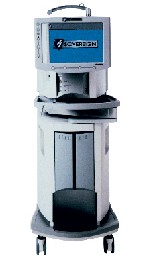 |
| The Sovereign (Advanced Medical Optics) is designed for bimanual phacoemulsification using a 1.2mm incision. |
 |
| The Infiniti (Alcon) offers three modes for cataract removal. |
 |
| The Millennium (Bausch & Lomb) offers Concentrix, which provides flow and vacuum response. |
Simultaneous Bilateral Cataract Surgery
Simultaneous bilateral cataract surgery (SBCS), also known as immediately sequential cataract surgery, is being performed on a limited basis outside the United States. However, the risk of SBCS is greater than potential benefits.4
Indeed, surgeons do not perform SBCS in the United States for several reasons, including:
Increased potential for lawsuits. One instance of bilateral endophthalmitis that results in significantly reduced visual acuity or bilateral retinal detachments could be grounds for an indefensible lawsuit.
Medicare regulations. Medicare penalizes the surgeon and comanaging optometrist 50% for a second procedure done in conjunction with the first.
Risk of IOL shifting. Surgically induced anisometropia, due to poor preoperative measurements or surgical techniques, can result in IOL shifting. This, in turn, causes unexpected refractive powers. This situation alone necessitates the removal of several IOLs each year.5
Perhaps the most comprehensive study examining the outcomes of SBCS was conducted by S. Arshinoff, who reported on 1,020 consecutive patients (2,040 eyes) who underwent bilateral surgery using either acrylic or silicone foldable IOLs.5 Thirty-two complications resulted from the procedures. Four patients suffered posterior capsule ruptures, one with vitreous loss. One patient suffered intraoperative hyphema when the cannula from the viscoelastic device came off and collided into the contralateral trabecula.
Another study emphasizes that bilateral corneal decompensation is a risk in SBCS.6 Interestingly, his research does not advocate routine preoperative corneal endothelial counts.
Other arguments against performing SBCS include bilateral protracted cystoid macular edema, ultrasound biometry accuracy within +/- 0.50D, ethical and financial considerations, and the fact that SBCS violates the 1996 American Academy of Ophthalmology Preferred Practice Patterns (PPO).6,7
Ultrasound vs. Er:YAG Laser
Ultrasound phacoemulsification, in which ultrasonic energy emulsifies cataracts of hardness, is the preferred surgical technique among cataract surgeons because of its effectiveness in removing soft- and medium-density cataracts.8
One major disadvantage, however: Increased water temperature (i.e., a temperature higher than normal eye temperature) can cause an inflammatory response and injury to the incision, intracameral structures, corneal endothelium, iris and posterior capsule.9
Another phacoemulsification energy source is sonic energy. The advantage of the erbium:YAG (Er:YAG) laser is its high level of water absorption. So, its laser beam can be focused directly on the cataract, while the high tissue water level shields adjacent tissues from injury.8 A downside, however, is that the incision size exceeds the 0.8mm that the laser allows.10
Researchers recently studied the effectiveness of infrared Er:YAG laser in removing nuclei of various densities.8 Specifically, they compared phacoemulsification to laser surgery. One study reported on 147 eyes (129 patients) that underwent standard ultrasound phacoemulsification or laser cataract surgery.8 The researchers classified each eye as 1+ (trace of nuclear sclerosis), 2+ and 3+ (moderate nuclear sclerosis), or 4+ (dense nuclear sclerosis). Also, they evaluated pachymetry, endothelial cells, IOP, uncorrected visual acuity, best-corrected visual acuity, corneal topography and fluorescein angiography.
The study concluded that the Er:YAG laser was as effective and reliable as ultrasound in removing soft- and medium-density cataracts. However, increased surgical length produced more trauma and complications, including significant endothelial cell loss, during laser treatment than during ultrasound treatment.
Surgeons in the United States and abroad have used this laser with varying degrees of success to remove hard and soft nuclei. For this reason, most cataract surgeons still prefer ultrasound phacoemulsification to laser cataract surgery.8
Because several types of IOLs are available, optometrists are beginning to recommend specific lenses to patients. Some of the newer options include:
Toric IOLs. These are now available from Staar Surgical and are made from their new collamer material. These lenses are available in cylinder powers of 2.00D and 3.50D, and they can be rotated 20 degrees from the horizontal axis. Patients with large amounts of corneal astigmatism may be candidates for this lens.
 |
| Toric IOLs (Staar Surgical) can be implanted in patients who have large amounts of corneal astigmatism. |
Array (Advanced Medical Optics). The Array offers multifocal vision to presbyopic patients who have cataracts. This lens is the only multifocal IOL currently approved by the Food and Drug Administra-tion. These lenses can provide multifunctional vision for patients who have moderate amounts of hyperopia, especially those with a loss of accommodation.
The Array has five zones that are alternately placed for near and far vision. If patients are properly selected (i.e., they are hyperopic and presbyopic with some cataract), they can enjoy far, intermediate and near vision. Some distance vision is sacrificed to gain increased near vision, however.
Crystalens (eyeonics, inc.). The crystalens is the first accommodating IOL to be approved by the FDA. The crystalens moves back and forth as a function of the movement of the vitreous face and works best in patients who have +1.00D to +3.00D of hyperopia.
 |
| The crystalens (eyeonics, inc.) is the first FDA-approved accommodating IOL. |
In the FDA clinical trial, 93.5% of subjects who were bilaterally implanted with the crystalens experienced visual acuity of 20/32 or better at near, intermediate or distance one year after surgery.11
Tecnis (Advanced Medical Optics). The Tecnis IOL is the first IOL to use wavefront technology. It has an anterior prolate surface that minimizes higher-order aberrations. This lens functions better under photopic and mesopic conditions than traditional acrylic lenses.12
SoFlex SE IOL (Bausch & Lomb). The SoFlex SE IOL, which is part of the SofPort system, has an aspheric anterior surface and, therefore, sharpens visual acuity similarly to the way aspheric contact lenses work. The SoFlex is a foldable silicone lens with a square edge to reduce posterior capsular opacification (PCO) formation.
IOL Design
We now know that round IOL edges allow migration of lens epithelial cells, which causes PCO and diminished vision.13 Therefore, YAG capsulotomy is often performed. Square-edged lenses, however, can cause unwanted optical images (i.e., dysphotopsia),13 which may prompt removal.
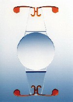
Manufacturers are looking for a compromise solution. To date, the OptiEdge (Advanced Medical Optics) appears promising. The OptiEdge features a square posterior edge to prevent lens epithelial cell migration, a round anterior edge, and a sloping intermediate portion to minimize unwanted internal reflections.
 |
| The OptiEdge (Advanced Medical Optics) features a square posterior edge, round anterior edge, and a sloping intermediate portion. |
Blue Light-Blocking Lenses
Ultraviolet (UV) light is often considered a cause of age-related macular degeneration (AMD), but data to support this theory is conflicting.14 Additionally, we do not yet know if the retina is normally exposed to damaging levels of blue light. Researchers are conducting investigations into whether blue light exposure in post-cataract patients can produce clinically significant retinal damage.
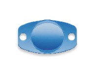
Concerns regarding the role of light in AMD led to the introduction of the AcrySof Natural IOL (Alcon Laboratories). Although all IOLs can block UV light, the AcrySof Natural blocks both UV and blue light.15 According to one study, a long-term population-based clinical trial would determine whether a blue light-absorbing IOL can reduce the risk for or progression of AMD.16
 |
| The AcrySof Natural (Alcon) filters ultraviolet and blue light. |
Cataract surgery and IOLs are continually evolving. Optometrists must keep up with these changes so they can make the best recommendations and provide the best care for their patients.
Dr. Smick is in private practice in Morrow, Georgia. He does not have a financial interest in any of the companies mentioned, but he is a consultant for Advanced Medical Optics.
1. Mamalis N. Is smaller better? J Cataract Refract Surg 2003 Jun;29(6):1049-50.
2. New strategies needed to reduce rate of post-cataract endophthalmitis. Primary Care Optometry News 2005 Feb 21.
3. Mino de Kaspar H, Chang RT, Singh K, et al. Prospective randomized comparison of 2 different methods of 5% povidone-iodine applications for anterior segment intraocular surgery. Arch Ophthalmol 2005 Feb;123(2):161-5.
4. Masket S. Consultation section: cataract surgical problem. J Cataract Refract Surg 1997 Dec;23(10):1437-41.
5. Arshinoff S, Strube YN, Tagev R. Simultaneous bilateral cataract surgery. J Cataract Refract Surg 2003 Jul;29(7):1281-91.
6. McDonnell PJ. Bilateral corneal decompensation after bilateral simultaneous cataract surgery. J Cataract Refract Surg 1999 Aug;25(8):1038.
7. Keskinbora HK. Simultaneous bilateral cataract surgery. J Cataract Refract Surg 1999 Mar;25(3):304-5.
8. Verges C, Llevat E. Laser cataract surgery: technique and clinical results. J Cataract Refract Surg 2003 Jul;29(7):1339-45.
9. Berger JW, Talamo JH, LaMarche KJ, et. al. Temperature measurements during phacoemulsification and erbium:YAG laser phacoablation in model systems. J Cataract Refract Surg 1996 Apr;22(3):372-8.
10. Agarwal S, Agarwal A, Sachdev MS, Mehta KR, eds. Laser phaco cataract surgery. In: Laser Cataract Surgery and Foldable IOLs. 2nd ed. New Delhi, India: Jaypee Brothers, 2000:237-46.
11. Food and Drug Administration. Crystalens model AT-45 accommodating posterior chamber intraocular lens (IOL) P030002. Available at http://www.fda.gov/cdrh/pdf3/
p030002.html. Accessed March 4, 2005.
12. Packer M, Fine H, Hoffman RS. Wavefront technology in cataract surgery. Curr Opin Ophthalmol 2004;15(1):56-60.
13. Smick KL, Masket S. New reflections in postoperative cataract care. Review of Optometry 2003;140(11):1CE-6CE.
14. Bressler NM, Bressler SB. Preventative ophthalmology: Age-related macular degeneration. Ophthalmology 1995;102(8):1206-11.
15. Alcon Laboratories. Open your eyes to the benefits of advanced cataract surgery. Available at http://cataractsurgery.com/us/patient/aboutlens.asp.
16. Sparrow JR, Miller AS, Zhou J. Blue light-absorbing intraocular lens and retinal pigment epithelium protection in vitro. J Cataract Refract Surg 2004 Apr;30(4):873-8.
