 History
A 35-year-old black female presented for a routine eye examination. She had no ocular complaints, signs or symptoms. Her systemic history was unremarkable. She reported no current medications and no known allergies.
Diagnostic Data
Her best-corrected visual acuity measured 20/20 O.U. at distance and near. The external examination was normal, with no evidence of afferent pupillary defect. Refraction revealed myopia consistent with her current spectacle correction.
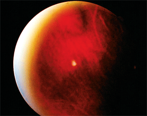
Her intraocular pressure was 19mm Hg O.U. The pertinent posterior segment findings are illustrated in the photograph.
Your Diagnosis
This patient presented
for a routine examination with no complaints or symptoms. However, we
noted an interesting lump on the anterior segment exam. Is this finding
normal?
How would you approach this case? Does this patient require any additional tests? What is your diagnosis? How would you manage this patient? What’s the likely prognosis?
Additional tests might include fundus photography to record the anomaly’s appearance and size; Amsler grid testing to rule out metamorphopsia or if any visual field is grossly missing; and automated field testing to quantify existing deficits. Because associated central nervous system arteriovenous malformations may be present, also consider retinology or neurosurgical referral to rule out the need for imaging, such as MRI or CT scan.
The diagnosis in this case is a vortex vein varix. The Latin word “varix” describes an enlarged and tortuous vein.1
Vortex veins are comprised of four to five large venous vessels located in loose connective tissue in the external vessel layer of the sclera.2 The vortex veins pierce the sclera to join the superior and inferior ophthalmic veins, which assist in carrying blood from the eye to the cavernous sinus.2
Varicies of the vortex vein ampullae are rare, benign, asymptomatic presentations that may be confused with choroidal nevi or melanomas.3 Initially, fluorescein angiography of the lesion indicates hypofluorescence, followed by isofluorescence 25 seconds after dye injection. Indocyanine green angiography often reveals a lesion that collapses when pressure is applied to the globe. If a choroidal mass collapses under applied pressure, you should suspect a vortex vein varix.
We have monitored this patient for the last five years. Her outcome was uneventful.
1. Friel JP. Varix. Dorland’s Medical Dictionary, 26th ed. Philadelphia: WB Saunders; 1985: 1437.
2. Snell RS, Lemp MA. The eyeball. In: Snell RS, Lemp MA (eds.). Clinical anatomy of the eye, 2nd ed. Malden, Mass.: Blackwell Science Inc.; 1998:132-213.
3. Kang HK, Beaumont PE, Chang AA. Indocyanine green angiographic features of varix of the vortex vein ampulla. Clin Experiment Ophthalmol. 2000 Aug;28(4):321-3.
