 History
A 22-year-old white female presented to the ophthalmology department following an emergency room visit with a chief complaint of worsening facial asymmetry. Her systemic and ocular histories were noncontibutory.
The patient reported no known allergies of any kind.
Diagnostic Data
Her best-uncorrected entering visual acuity measured 20/20 OU at distance and near. There was no evidence of afferent pupillary defect or visual field involvement OU. The anterior segment findings were normal in both eyes.
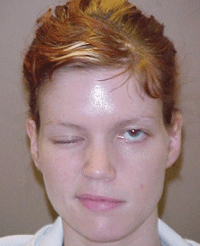
Intraocular pressure measured 14mm Hg OU. Dilated fundoscopy was within normal limits in both eyes. The external examination findings are illustrated in the photograph.
Your Diagnosis
How would you approach this case? Does the patient require any additional tests? What is your diagnosis? How would you manage this patient? What is the likely prognosis?
Discussion
Further testing included additional patient history regarding recent wind or cold exposure on the face. We also asked her about any recent upper respiratory illnesses.
In an effort to determine the type and extent of facial nerve injury, we instructed the patient to smile. Additionally, we tested her lid closure capacity, and determined that she had a grade IV lagophthalmos OS.
Further, we evaluated her corneal sensitivity to rule out the possibility of damaged sensory nerves. We used sodium fluorescein dye to assess the magnitude of the epitheliopathy.

|

|
|
Gross external images of our 22-year-old patient revealed obvious facial asymmetry. What is the correct diagnosis, and how should she be managed?
| |
The diagnosis in this case is cranial nerve VII (CN VII) palsy secondary to developing Guillain-Barre syndrome (GBS).
The division of the seventh cranial nerve is responsible for delivering the voluntary motor innervation to the muscles of facial expression, as well as to the stapedius muscle of the inner ear to help dampen loud sounds.1-6
The orbicularis oculi is responsible for eyelid closure and is under the control of CN VII.1-6 Consequently, damage to one CN VII’s nuclei or fascicles, or interruption to its peripheral course, will cause weakness or paralysis of one side of the face with an inability to voluntarily close the ipsilateral eye. Additional findings (seen on the affected side) include nasal labial fold flattening, mouth corner droop, ectropion, lagophthalmos, decreased tear production, tear deficiency and evaporative dry eye, conjunctival injection, corneal compromise, decreased taste from the anterior two thirds of the tongue and hyperacusis (supersensitivity to sound).1-5
The muscles that control the voluntary responses of smiling, frowning, grimacing and eyelid closure depend on signals that originate in the facial motor area of the precentral gyrus associated with the frontal cerebral cortex.2,3 Supranuclear motor neurons connecting cortical areas 4 and 6 with the facial nuclei descend as fascicles of the corticobulbar tract through the internal capsule to the level of the lower pons via the cerebral peduncles.2,3 The portion of each facial nucleus that controls the muscles of the upper face receives corticobulbar stimulation from both the right and left precentral motor corticies.
The supranuclear innervation supporting the muscles of facial expression in the lower face is crossed only.2,3,5 The muscles that close the eyes and wrinkle the forehead are bilaterally innervated; therefore, a lesion in the cortex or supranuclear pathway on one side spares eyelid closure and forehead wrinkling, but results in contralateral paralysis of the lower face.2,3
Because the area of the cortex associated with facial muscle function lies near the motor representation of the hand and tongue, weakness of the thumb, fingers and tongue located ipsilateral to the facial palsy may occur.2-5 Also, because supranuclear lesions produce spastic, rather than flaccid, paralysis. This permits less significant nasolabial fold and mouth corner droop than its lower motor neuron counterpart.2,3
The facial motor nuclei are located in the lower pontine tegmentum and have an intimate relationship with the trigeminal nerve (CN V), abducens nuclei (CN VI), cochlear nuclei (CN VIII), the medial longitudinal fasciculus, the paramedian pontine reticular formation (PPRF), the descending coticospinal fibers and descending sympathetic fibers.
The facial nucleus contains four separate cell groups that innervate specific muscle groups. Motor axons exit the nucleus dorsally, loop around the CN VI nuclei and emerge into the subarachnoid space from the lateral aspect of the pons.2,3,5 Fibers from the superior salivary and lacrimal nuclei join the facial nerve at the cerebellopontine angle. CN VIII is present here, as well.
Lesions at this level include temporal bone fractures and infections, schwannomas, neuromas (cerebellopontine angle tumors) and vascular compressions, and may produce deficits in hearing, balance, tear production and salivary flow.2,3,5
The facial (CN VII) and the vestibuloacoustic nerves enter the internal auditory meatus together.1-3 The facial nerve then departs from the acoustic nerve to enter the fallopian (facial) canal, which courses 30mm through the temporal bone and incorporates the geniculate ganglion.2
Lesions that involve the ganglion include geniculate ganglionitis. Acoustic neuroma that involve CN VIII can impair hearing, facial nerve function and produce corneal hypoesthesia (CN V). Lesions that begin within the nucleous or along the fascicles are believed to involve the final common pathway of neural transmission, and are known as lower motor neuron or peripheral lesions.
The first major branch of CN VII, the greater superficial petrosal nerve, traverses the geniculate ganglion, proceeds forward, crosses the dura mater of the middle cranial fossa and synapses in the sphenopalatine ganglion. The sphenopalatine ganglion gives rise to postganglionic fibers that join the zygomatic and lacrimal nerves of CN V to innervate the lacrimal gland. Lesions here impair reflex tear secretion. It is important to note, when defective tear production accompanies CN V or CN VI palsy, middle cranial fossa disease is indicated.2,3,5
The stapedius branch of CN VII arises from the distal segment of the facial nerve.2,3 Lesions found here disable the patient’s ability to dampen sound, producing hyperacusis. As the facial nerve continues downward, the chorda tympani branch arises from the canal. The chorda tympani contains sensory afferent fibers that transmit taste sensation from the anterior two-thirds of the tongue. It also contains autonomic (parasympathetic preganglionic) nerve fibers that innervate the submandibular and sublingual salivary glands.2,3
Lesions located anywhere along this pathway cause an interruption in salivary flow, as well as inhibit taste receptor function in the anterior two-thirds of the tongue.2,3 The portion of the facial nerve that contains the motor fibers which innervate the muscles of facial expression exits the stylomastoid foramen and enters the substance of the parotid gland before distribution.2,3
Therefore, lesions of the parotid gland also must be investigated as part of the diagnostic work-up. Sensory afferents from the external auditory meatus and a small area of skin behind the ear transmit pain, temperature and touch information.2,3
The most common cause of unilateral facial weakness is idiopathic (53%).2 These lesions are theorized to occur secondary to idiopathic inflammation, viral infection or vascular compression of CN VII. Given the extensive neurology of CN VII, idiopathic “Bell’s Palsy” is a diagnosis of exclusion.
The most common causes of peripheral CN VII palsy include cerebellopontine angle tumor, trauma, otitis media, herpes zoster oticus, Lyme disease, sarcoidosis, GBS, Epstein-Barr virus, parotid neoplasm, syphilis, diabetes mellitus, herpes simplex, herpes zoster, pregnancy and HIV.1-11
Guillain-Barre syndrome is an acute, demyelinating polyneuropathy involving the spinal roots, peripheral nerves and often the cranial nerves. Although its exact mechanism remains unclear, an autoimmune etiopathology is theorized. It is characterized by rapidly progressing, symmetrical muscular weakness starting in the legs and ascending to the trunk and arms. Additionally, affected individuals experience compromised deep tendon reflexes.
Approximately half of the patients with GBS develop cranial nerve palsies, with unilateral or bilateral facial nerve (CN VII) palsy being the most common. Paralysis of the tongue, lips, palate, larynx and pharynx muscles secondary to lesions involving cranial nerves V, IX, X and XI are the next most common nerve-related abnormalities. Ocular muscle palsy is not common, occurring in just 10% of patients. The Miller Fisher variant of GBS is a distinct syndrome in which the only neurologic deficits are oculomotor palsies, areflexia and ataxia.12-17
In acute GBS, the myelin sheaths of the peripheral nerves and nerve roots become infiltrated with lymphocytes and macrophages. Subsequently, the myelin sheaths and their progenitors, the Schwann cells, undergo segmental destruction from a lymphocyte-mediated autoimmune reaction.12-14,16-18 The pathology results in degradation of the rapid, saltatory neural conduction that is characteristic of myelinated nerve fibers.12-14,16,17 In severe lesions, axonal degeneration also occurs. Further, in a few patients, the primary abnormality is acute axonal degeneration, rather than demyelination.12-14,16,17
The optometric management of a patient who presents with CN VII palsy begins with obtaining a complete history, performing a cursory evaluation of the 12 cranial nerves and executing a comprehensive ocular examination with dilated fundus and optic nerve evaluation.
Close attention should be given to the affected eyelid’s posture, corneal wetting (tear film break-up time), blink posture, tear quality (sodium fluorescein staining) and tear quantity (Schirmer tear testing). In cases where the diagnosis is questionable (upper motor neuron lesion), instruct the patient to close both eyes, and then apply force to attempt lid opening. If one lid is significantly easier to open than the other, suspicion should be raised.
Exposure keratopathy can be managed with ocular lubricating drops and ointments. Moisture chamber patches or eyelid taping also are possible solutions. Moisture chamber patches can be attached to spectacle temples to create a dampened ocular environment and lessen tear evaporation. Temporary external eyelid weights also have been described as devices that can bridge the time span between paralysis onset and a determination of surgical nescessity.7
Because idiopathic facial nerve palsy is a diagnosis of exclusion, laboratory testing (e.g., Lyme titre, rheumatoid factor, erythrocyte sedimentation rate, antinuclear antibody, echocardiogram, fluorescent treponemal antibody absorption test, HIV titre, chest x-ray), lumbar puncture (in patients with suspected neoplasm), neuroradiologic studies (computed tomography and magnetic resonance imaging) and appropriate referrals (otolaryngology, neurology and neurosurgery) should be obtained.1-6
Treatment for peripheral facial weakness depends upon the etiology, and usually is placed under the specialist’s discretion.4 According to one study, most of the available evidence from randomized controlled trials show no significant or clear benefit to treating Bell’s palsy with systemic corticosteroids.9 Another research team asserts the same position for oral antivirals.10 Because use of these agents rarely creates complications, their introduction remains a matter of individualized management preference.
GBS is a medical emergency, requiring constant monitoring and support of vital functions.13-16 The airway must be kept clear, and respiration can be assisted if necessary. Additionally, fluid intake should be sufficient to maintain a urine volume of at least 1L to 1.5L per day.
In GBS, corticosteroids often worsen the outcome and should not be used.13-15 Plasmapheresis helps when performed early in the disease, is relatively safe, shortens the disease course and hospitalization, and reduces mortality and the incidence of permanent paralysis. More importantly, it is the treatment of choice for acutely ill GBS patients.
Daily IV infusion of 400mg/kg/day of immune globulin for the first two weeks following GBS diagnosis is as effective as plasmapheresis, and may be safer. Therefore, if the patient does not respond to plasmapheresis, it is unavailable or it cannot be performed (due to difficult venous access or hemodynamic instability), globulin should be used.13-15,19-21
It has also been shown that sequential treatment of plasmapheresis followed by globulin treatment, or vice versa, does not have an increased effect compared to monotherapy.
Thus, sequential treatment is not recommended.22 Immunomodulator therapy, although helpful in multiple sclerosis, is not an effective management approach. In fact, some researchers believed that interferon-α may even trigger the development of GBS.22
In chronic relapsing polyneuropathy, corticosteroids improve weakness and may be required over the long-term. Immunosuppressive drugs, such as azathioprine, may benefit some patients.13,14
As for out patient, we managed her Bell’s palsy with a combination of manual blinking, artificial drops and ointments, and moisture-chamber patches. We offered the patient tarrsoraphy, but she refused.
She underwent systemic treatment with a combination of IV corticostreroids and immunoglobulin infusions. She recovered completely over a four-month period, and showed no evidence of residual cosmetic issues.
1. Gurwood AS. The Eyelid and Neuro-ocular Disease. In: Blaustein BH. Ocular Manifestations of Neurologic Disease. Philadelphia: Mosby; 1996:127-51.
2. Gurwood AS, Tasca JM, Kulback E. A review of cranial nerve VII palsy with emphasis on Bell’s palsy. South J Optom. 1996;14(3):13-7.
3. May M, Galetta S. The Facial Nerve and Related Disorders of The Face. In: Glaser JS. Neurophthalmology, 2nd ed. Philadelphia: J.B. Lippincott Co.; 1990:239-77.
4. Savino PJ, Sergot, RC. Neuroophthalmology: Isolated Seventh––Nerve Palsy. In: Rhee DJ, Pyfer MF. The Wills Eye Manual, 3rd Ed. Philadelphia: Lippincott Williams and Wilkins; 1999:290-4.
5. Bajandas FJ, Kline LB. Seven Syndrome of The Seventh (Facial) Nerve. In: Bajandas FJ, Kline LB. Neuro-ophthalmology Review Manual, 3rd ed. Thorofare, NJ.: Slack Inc; 1988:151-6.
6. Danielides V1, Skevas A, van Cauwenberge P, et al. Facial nerve palsy during pregnancy. Acta Otorhinolaryngol Belg. 1996;50(2):131-5.
7. Zwick OM, Seiff SR. Supportive care of facial nerve palsy with temporary external eyelid weights. Optometry. 2006 Jul;77(7):340-2.
8. Sensat ML. Mobius syndrome: a dental hygiene case study and review of the literature. Int J Dent Hyg. 2003 Feb;1(1):62-7.
9. Salinas RA, Alvarez G, Ferreira J. Corticosteroids for Bell’s palsy (idiopathic facial paralysis). Cochrane Database Syst Rev. 2010 Mar 17;(3):CD001942. doi: 10.1002/14651858.CD001942.pub4.
10. Allen D, Dunn L. Aciclovir or valaciclovir for Bell’s palsy (idiopathic facial paralysis). Cochrane Database Syst Rev. 2004;(3):CD001869.
11. Gilbert SC. Bell’s palsy and herpesviruses. Herpes. 2002 Dec;9(3):70-3.
12. Shier D. The Nervous System. In: Shier D, Butler J, Lewis R. Hole’s Textbook of Anatomy and Physiology, 9th Ed. New York: Mcgraw-Hill; 2002:215-51.
13. Fine EJ, Selman WR. Neurologic Disorders: Spinal Cord Disorders. In: Beers MH, Berkow R. The Electronic Merck Manual. Available at: www.merck.com/pubs/mmanual/section14/chapter182/182g.htm. Accessed June 2, 2014.
14. Fine EJ, Selman WR. Neurologic Disorders: Spinal Cord Disorders. In: Beers MH, Berkow R. The Electronic Merck Manual. Available at: www.merck.com/pubs/mmanual/section14/chapter183/183f.htm. Accessed June 2, 2014.
15. Susuki K, Johkura K, Yuki N, et al. Rapid resolution of nerve conduction blocks after plasmapheresis in Guillain-Barre syndrome associated with anti-GM1b IgG antibody. J Neurol. 2001 Feb;248(2):148-50.
16. Hadden RD, Karch H, Hartung HP, et al. Preceding infections, immune factors, and outcome in Guillain-Barrésyndrome. Neurology. 2001 Mar 27;56(6):758-65.
17. Kuwabara S, Mori M, Ogawara K, et al. Indicators of rapid clinical recovery in Guillain-Barré syndrome. J Neurol Neurosurg Psychiatry. 2001 Apr;70(4):560-2.
18. Glocker FX, Rösler KM, Linden D, et al. Facial nerve dysfunction in hereditary motor and sensory neuropathy type Iand III. Muscle Nerve. 1999 Sep;22(9):1201-8.
19. Yuki N, Odaka M, Hirata K. Acute ophthalmoparesis (without ataxia) associated with anti-GQ1b IgGantibody: clinical features. Ophthalmology. 2001 Jan;108(1):196-200.
20. Finsterer J. Treatment of immune-mediated, gysimmune neuropathies. Acta Neurol Scand. 2005 Aug;112(2):115-25.
21. Newswanger DL, Warren CR. Guillain-Barré syndrome. Am Fam Physician. 2004 May 15;69(10):2405-10.
22. Hughes RA, Wijdicks EF, Barohn R, et al. Practice parameter: immunotherapy for Guillain-Barré syndrome: report of the Quality Standards Subcommittee of the American Academy of Neurology. Neurology. 2003 Sep 23;61(6):736-40.

