Recent research has demonstrated the important influence of peripheral defocus on eye growth, as well as labeled peripheral hyperopia defocus as a high-risk factor for the development and progression of myopia. A new study sought to investigate peripheral refractive status in children with varying refractive states using a novel imaging technology called multispectral refractive topography (MRT), which allows for peripheral refraction of the different regions of the retina. A main takeaway from the results, published recently in the Journal of Ophthalmology, is that peripheral defocus has significant implications for the genesis of myopia.
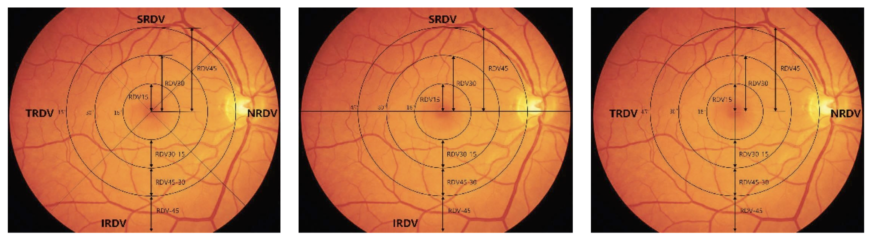 |
| Multispectral refractive topography may be used in the future as an objective method to assess peripheral refraction in multiple discrete regions in patients with or at risk for developing myopia. Photo: Zhao Q, et al. J Ophthalmol. August 8, 2024. Click image to enlarge. |
Included in the study were 814 subjects (814 eyes) divided into three groups according to the central spherical equivalent refraction (SER): emmetropia group, low myopia group and moderate myopia group. The researchers used MRT to measure the retinal absolute and relative refractive difference value (RDV) in four different regions—superior, inferior, temporal and nasal—defined based on several concentric circles extending outward from the macular fovea.
Analysis of the data showed that in all regions, the absolute value of RDV decreased with increasing degree of myopia. Consequently, eyes with different refractive degrees had different relative values of RDV. One example the authors gave in their paper was that “in nasal position within 45° and temporal position within 30°, the peripheral retina exhibited significantly different relative hyperopic refractive status” among the emmetropia, low myopia and moderate myopia groups.
The researchers also examined the potential correlations between SER and axial length and RDV. They found that SER was negatively correlated with nasal RDV within 30°, positively correlated with temporal RDV within 15° and not significantly correlated with superior RDV and inferior RDV when the retina was divided into four parts. As for axial length, researchers found a positive correlation with nasal RDV within 30° and a negative correlation with temporal RDV within 15°.
“The peripheral defocus of the horizontal direction has a greater effect on the development of myopia in children,” the researchers concluded in their paper. “Moreover, RDV within the range of nasal RDV-30 is most closely associated with the development of myopia.”
While adding to the evidence of a relationship between peripheral defocus and myopia development, this study also demonstrates MRT as a reliable tool to assess peripheral retinal refraction. “Autorefractometers are currently the gold standard for testing refraction of the central retina,” the study authors wrote. “Liao et al. proved that autorefractometry and MRT show high agreement in measuring central refraction, and MRT could provide a potential objective method to assess peripheral refraction. Hence,” they noted, “the advantages of MRT can be used to solve more problems for myopia in the future.”
Zhao Q, Wang Y, Liang T, Nie W, Xue P, Cheng J. Characteristics of peripheral retinal refraction and its role in children with different refractive states. J Ophthalmol. August 8, 2024. [Epub ahead of print]. |