 |
|
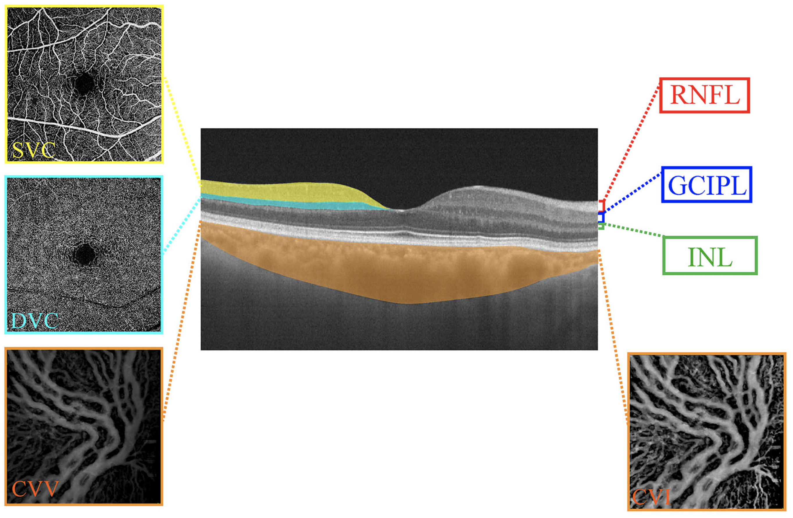
This analysis revealed a rapid and significant increase in superficial vascular complex density and choroidal vascular volume in ipsilateral eyes following carotid artery stenting. Photo: Cao L, et al. Transl Vis Sci Technol. August 2, 2024. Click image to enlarge. |
New findings suggest that OCT and OCT angiography (OCT-A) have the potential to detect changes in the retina and choroid following carotid artery stenting in patients with unilateral stenosis. These findings—recently published in the journal Translational Vision Science & Technology—could guide future research on the long-term effects of stenting on these ocular structures.
In this analysis, which is part of an ongoing cohort study, researchers used OCT-A to evaluate the effect of carotid artery stenting on the retina and choroid. Baseline enrollment included 303 eyes from 160 patients.
The investigators imaged all study eyes and evaluated various OCT/OCT-A metrics, including retinal structural thicknesses and microvasculature and choroidal parameters (choroidal vascular volume/index), before carotid artery stenting as well as four days and three months after the procedure.
Data showed that superficial vascular complex (SVC) and deep vascular complex (DVC) densities as well as choroidal vascular volume (CVV) were lower in ipsilateral eyes (stenosed side) vs. contralateral eyes. Four days following stenting, researchers observed a significant increase in SVC density in ipsilateral eyes while a significant increase was seen in CVV among ipsilateral eyes and contralateral eyes.
Additionally, the researchers reported a significant decrease three months after stenting (63 patients with 114 eyes) in the ganglion cell–inner plexiform layer (GCIPL) thickness of ipsilateral and contralateral eyes.
Use of swept-source OCT “enables a fast-scanning speed, permitting denser scan patterns and larger scans,” the study authors noted in their TVST paper. “Moreover, the tool operates with a longer wavelength, facilitating enhanced light penetration through the retina and choroid, which may help resolve the controversies surrounding the effect of carotid stenting on the eye.”
Findings from this investigation showed that four days after stenting, RNFL thickness, SVC density and CVV increased while GCIPL thickness decreased, the research team summarized, while also highlighting the need for further study. “The long-term effect of carotid stenting on the retina and choroid should be explored by longer follow-up.”
Cao L, Wu J, Wang H, et al. Influence of carotid artery stenting on the retina and choroid. Transl Vis Sci Technol. August 2, 2024 [Epub ahead of print]. |