Findings from a recent analysis, published in the journal Ophthalmic and Physiological Optics, highlight the additional detail afforded by incorporating anterior segment OCT imaging in addition to slit lamp imaging to enhance the accuracy of cataract assessment.
This study, which evaluated whether adding OCT imaging of the crystalline lens to standard clinical assessments improves the accuracy and precision of lens opacity evaluation as well as the clinical management decisions of optometrists, included 50 ODs actively working in the clinical setting. Thirty-six reported working in primary eye care and 14 in hospital settings.
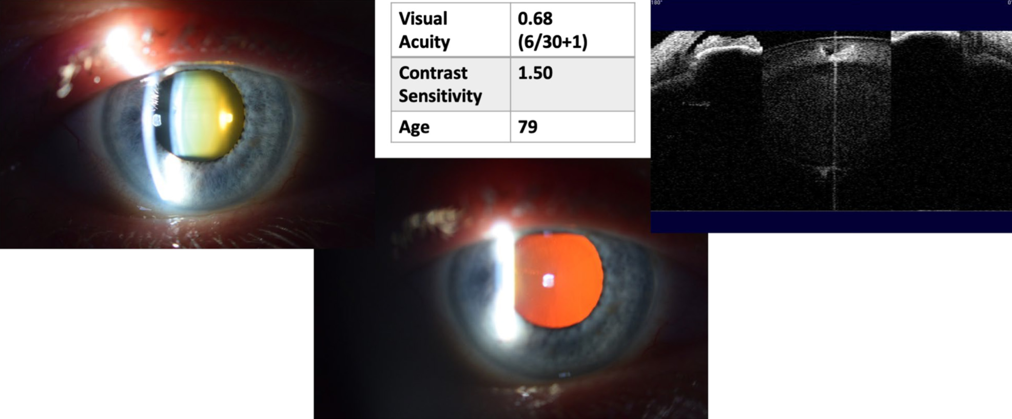 |
| New research supports the notion of considering anterior segment OCT as an adjunct to slit-lamp images to help improve the accuracy of cataract assessment. This image from the study shows slit lamp images, OCT lens section and corresponding visual function measures. Photo: Burke N, et al. Ophthalmic Physiol Opt. Click image to enlarge. |
“Clinical vignettes were developed from anonymized data sets of 50 individuals collated during previous research studies where participants with varying severity of lens opacity were recruited,” according to the study authors. There were three forms of vignettes: slit-lamp images of the lens, slit-lamp and OCT images, and slit-lamp, OCT and visual function measures.
All participating optometrists completed the study. Data showed that agreement was highest where vignettes contained all clinical data (i.e., slit lamp, OCT and visual function data—grading: 51.0%, management: 50.5%) and systematically reduced with decreasing vignette content, the investigators reported.
In their paper, they also noted that, “A larger number of vignettes containing imaging and visual function measures exhibited below reference (i.e., less conservative) grading compared with vignettes containing imaging data alone.”
The analysis revealed that optometrists based in the hospital setting were less conservative in grading lens opacities when compared to their counterparts in primary eyecare. In terms of years of experience, the researchers observed no significant association with grading decisions, suggesting that this did not influence the accuracy of cataract assessment.
“This study found that optometrist's precision of grading improved when more clinical information was given, and that OCT imaging compared with the slit-lamp alone increased the proportion of accurate grading assessments,” the study authors noted in their Ophthalmic and Physiological Optics paper. “However, structural assessment alone yielded more conservative decision making, which reversed when visual functional data was available.
As we seek to stratify and improve cataract referral pathways, inclusion of anterior segment OCT may improve the precision of cataract grading,” they concluded.
Burke N, Mulholland PJ, Keane PA, et al. Investigating the impact of OCT imaging of the crystalline lens on the accuracy and precision of cataract assessment. Ophthalmic Physiol Opt. August 23, 2024 [Epub ahead of print]. |