 |
|
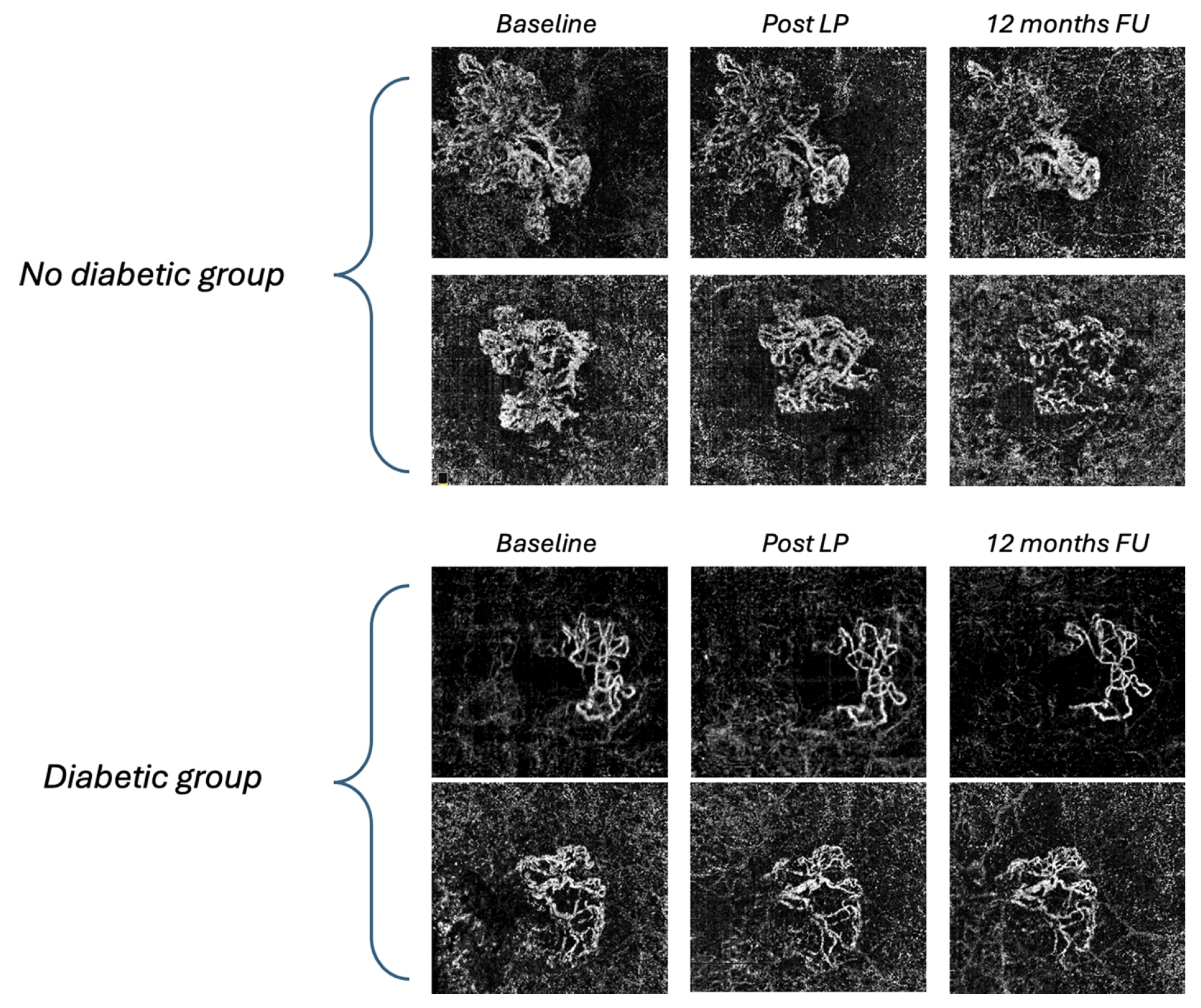
In a recent study published in the journal Investigative Ophthalmology & Visual Science, researchers followed two groups of patients with nAMD (one with mild DR, and the other without) who received anti-VEGF therapy for one year. Their most significant discovery was in the size of macular neovascularization lesion size, which continued to decrease in the non-diabetic group over the course of 12 months, but had a smaller decrease in the diabetic group, suggesting the disease may be a risk factor to consider during anti-angiogenic treatment. Photo: Boscia G, et al. Invest Ophthalmol Vis Sci. August 2, 2024. Click image to enlarge. |
Several environmental and genetic factors contribute to the pathogenesis of age-related macular degeneration (AMD), but many in the scientific community debate the role of diabetes mellitus (DM) in the more advanced neovascular AMD (nAMD) questioning whether it contributes to or protects against the disease progression. Using OCT-A to observe the morphological changes in type 1 exudative macular neovascularization (MNV) following one year of anti-VEGF therapy, researchers found patients with mild diabetic retinopathy (DR) had a divergent response in type 1 MNV lesion area.
According to their findings, recently published in the journal Investigative Ophthalmology & Visual Science, although there was a significant reduction in lesion size between the baseline visit and the post-loading phase, there was no noticeable further reduction in lesion area at the 12-month follow-up visit. Alternatively, nAMD patients with no history of DM had a continuous reduction in MNV size. Researchers say this highlights the significance of DR as a potential modifier of treatment outcomes in nAMD management, with DM considered a risk factor during anti-angiogenic treatment.
The retrospective study included 45 eyes with exudative nAMD with type 1 MNV, all of whom were enrolled at the Medical Retina Service at the University of Bari Aldo Moro in Italy. Patients were divided into the Diabetic group, which included 21 eyes of 21 patients with mild DR; and Not Diabetic Group, which consisted of 24 eyes of 24 patients with no history of DM. The outcome measures included best corrected visual acuity changes, central macular thickness, MNV lesion area, and MNV flow area. All of these parameters showed significant improvement after the loading phase (LP), according to the study.
“The most interesting finding pertained to the behavior of the neovascular lesion after 1 year of treatment initiation,” they wrote in the study. “Specifically, the Diabetic group did not exhibit a reduction in MNV after 12 months; instead, there was a lack of significant reduction of the area of the MNV. In contrast, the Not Diabetic group showed a continuous reduction in the size of the MNV. This last result appears to be highly significant, especially considering that the size of MNV evaluated with OCT-A serves as a valuable biomarker in assessing the response or lack of response to treatment.”
They speculated about the mechanisms that could be contributing to this result, including the possibility that the presence of DR alters the angiogenic pathways involved in MNV. “DR is associated with dysregulated VEGF signaling and increased levels of inflammatory cytokines, which could potentially lead to a different response to anti-VEGF therapy,” they said. “These altered pathways may result in a reduced responsiveness to anti-VEGF therapy, leading to a less pronounced reduction of neovascularization over time. Another possibility is that the microvascular changes associated with DR, such as capillary dropout and basement membrane thickening, create a less conducive environment for the resolution of neovascular complexes. The compromised vascular structure and function in diabetic eyes may impede the ability of anti-VEGF therapy to induce significant regression of the MNV, resulting in a lesser degree of reduction compared to non-diabetic eyes.”
The sample size was relatively small, noted the authors, which could have potentially limited the generalizability of their results. The lack of control group also means observed differences during the follow-up period could have occurred even without anti-angiogenic therapy. “Furthermore, the use of spectral-domain OCT-A, which utilizes shorter wavelength light compared to swept-source OCTA, may result in reduced signal penetration through the RPE, potentially affecting the accuracy of our imaging data,” they added.
Despite these factors, the authors say they felt confident in the strengths of their study, and say it’s the first comprehensive investigation of the impact of DR on longitudinal morphological and functional changes in type 1 MNV associated with AMD in patients undergoing anti-VEGF therapy for 1 year. The divergent response they discovered in this study highlights the role DR plays in treatment outcomes, and they concluded saying, “Future larger studies utilizing swept-source OCT-A and longer follow-up durations are necessary to validate our preliminary findings.”
Boscia G, Bacherini D, Vujosevic S, Grassi MO, Borrelli E, Giancipoli E, Landini L, Pignataro M, Alessio G, Boscia F, Viggiano P. Long-term impact of diabetic retinopathy on response to anti-VEGF treatment in neovascular AMD. Invest Ophthalmol Vis Sci. August 2, 2024. [Epub ahead of print.] |