Using widefield swept-source OCT angiography, researchers in China wanted to investigate the effects of refractive error on the inner layers of the retina. Specifically, they were interested in potential ganglion cell complex thickness (GCCT) and retinal capillary density (CD) effects caused by high myopia.
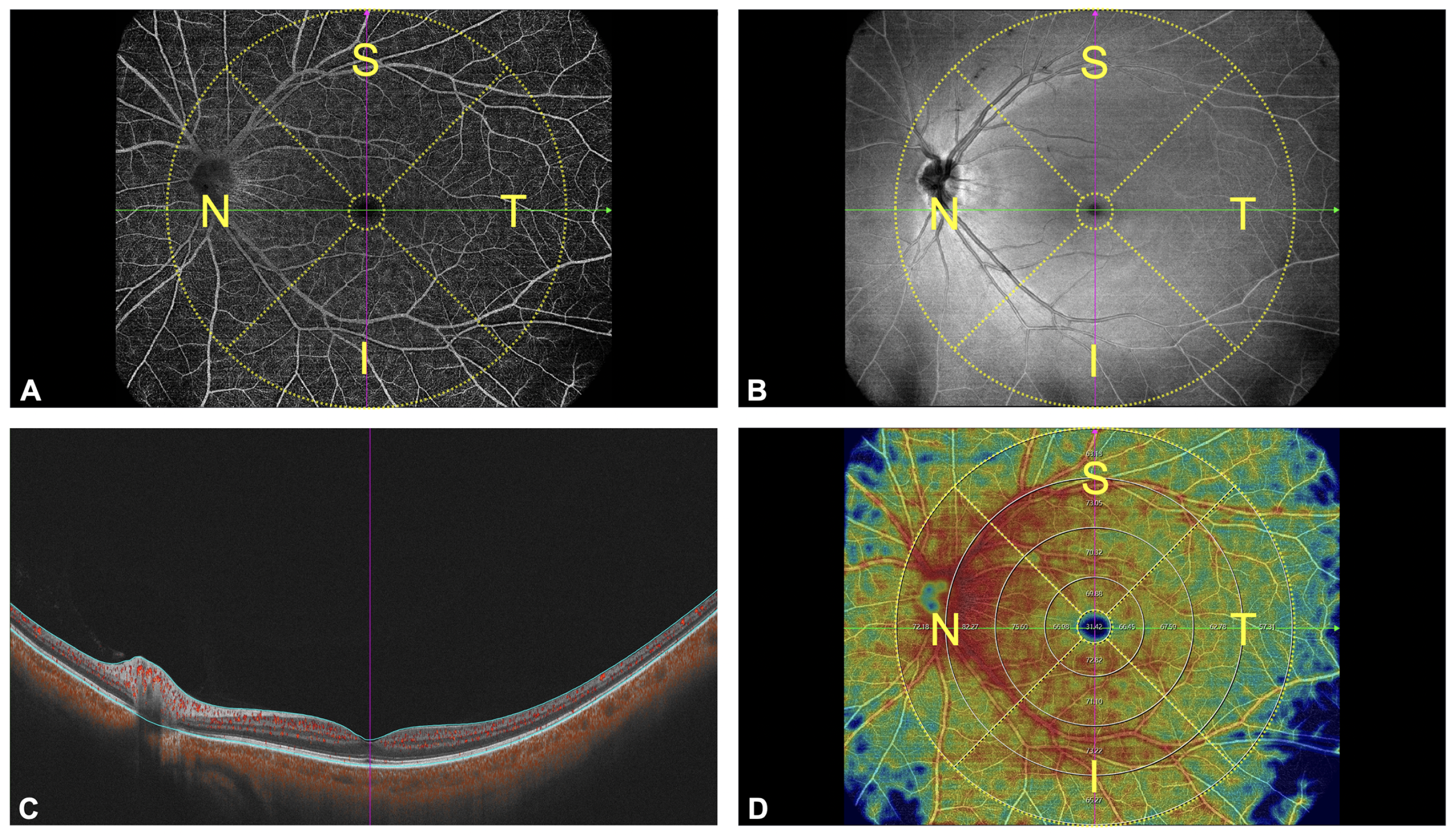 |
| The authors speculate the reason that retinal microvasculature might be more affected by axial elongation on the temporal side is because of larger vessel presence in proximity to the nasal side of the optic disc. This image from the study shows (A) a 15x12mm2 widefield OCT-A scan, (B) the corresponding en-face image, (C) a B-scan image and (D) measurement of the capillary density in annular regions of 1-3mm, 3-6mm, 6-9mm and 9-12mm in diameter, which were further divided into four quadrants(inferior, nasal, superior, temporal). Photo: Wang D, et al. Transl Vis Sci Technol. 2024;13(8):4. Click image to enlarge. |
The retrospective design included 506 total eyes; fovea-centered scans were obtained to assess subregional GCCT and CD across the entire retina, the superficial capillary plexus (SCP) and deep capillary plexus (DCP) in three different groups: a control group, a high myopic group of eyes with axial length <28mm and another highly myopic group but with eyes >28mm of axial length.
Their data showed that in myopic eyes with >28mm axial length, GCCT and retinal CD displayed a general decline in most regions. In myopic eyes with axial length <28mm, great reductions were seen in peripheral regions, including the GCCT beyond the 3x3mm2, CD in the 9mm to 12mm whole retina, 9mm to 12mm superior SCP as well as in 6mm to 12mm DCP. Maximum GCCT and retinal CD reduction with axial elongation was seen in subregions beyong 6x6mm2.
From these data, the authors of the study suggest that “the peripheral retinal GCC and microvasculature are more susceptible to the effects of axial elongation” and add that, “to our knowledge, this is the first study to report these findings, highlighting the differential impact of axial elongation on the peripheral retinal structures across varying degrees of high myopia.”
The authors’ discussion elaborates on these results. This is not the first study to demonstrate that high myopia can affect the retina, as previous reports indicate it can cause macular retinal microvasculature reduction. The current study demonstrated a preferential effect of axial elongation of CD in the temporal DCP; as more evidence has come out, this may be due to the DCP being more susceptible to high myopia effects than the SCP because of mechanical stretching from axial hyperextension in highly myopic eyes, as well as smaller-diameter, vulnerable vessels in the DCP more likely to be straightened and disrupted than larger retinal vessels of the SCP.
The investigators also point out that GCC thinning in most regions of this study correlated with increasing myopia, suggesting that the GCCT beyond the 3x3mm2 area decreased more significantly during myopia progression. Another important finding was that only myopic eyes with axial length >28mm displayed significant decrease in overall SCP CD.
Because of this sort of discrepancy compared with results in myopic eyes with less axial elongation, the researchers relay that, “therefore, it is necessary to evaluate different regions during the clinical assessment of the effect of myopia on the fundus and pay close attention to the peripheral retina.”
Additionally, they hope that “our study will contribute to understanding the role of the peripheral retinal microvasculature in the pathophysiological mechanisms of myopia and facilitate further advances in the clinical management of myopia.”
Wang D, Zhang Y, Lin F. Peripheral ganglion cell complex thickness and retinal microvasculature in myopia using wide-field swept-source OCT. Transl Vis Sci Technol. 2024;13(8):4. |