 |
|
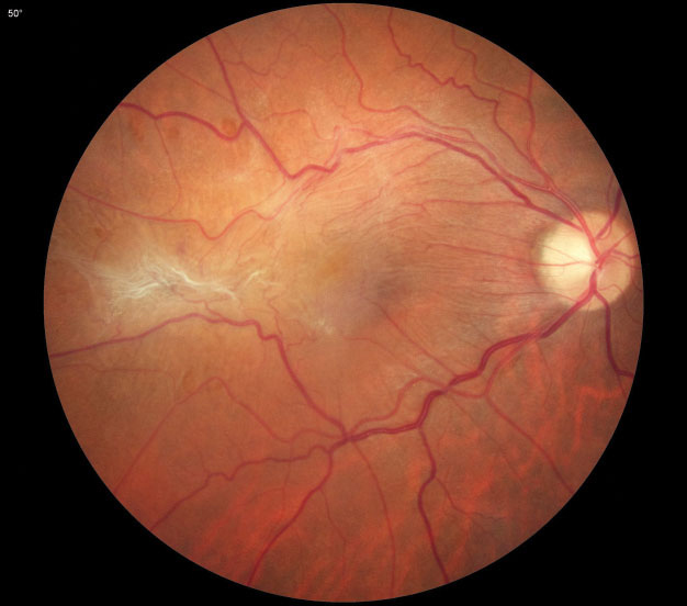
Epiretinal membrane and rhegmatogenous retinal detachment are both associated with metamorphopsia. ERM contraction may compress photoreceptor distribution in the macula, causing macropsia, and macula-off retinal detachments resulting in photoreceptor separation have been associated with micropsia. Photo: Diana Shechtman, OD. Click image to enlarge. |
It’s tough to know exactly what a patient sees, particularly if they have a visual disorder such as metamorphopsia. The symptom, often arising from displaced photoreceptors at the macula, is commonly present in AMD and vitreoretinal conditions; understanding the patient’s experience of it can be valuable to disease management. The tried-and-true Amsler grid isn’t able to map the exact image distortion experience in any way that a clinician can experience first-hand. Recently, however, researchers from London published a novel method of assessing this visual phenomenon called the Image Warping Test (IWT). Their paper, published in Retina, found that this test compared favorably to established measurement tools.
The IWT is a uniocular test in which the patient is presented with a grid of black lines on a white background displayed on a computer monitor. This background is dynamically warped by an operator until the grid lines appear straight to the patient and the metamorphopsia is cancelled out. “The reversal of this correction generates a grid which represents the pattern of the participant’s metamorphopsia,” the researchers explained in their paper.
They analyzed metamorphopsia measurements of 25 volunteer subjects who had metamorphopsia secondary to vitreoretinal pathology. Tests included standard Amsler grid, a modified Amsler grid called Morphision (in which the central area is presented in a warped pattern), the M-Charts collection of grids and the IWT.
They reported no correlation between best-corrected distance visual acuity and IWT score or between M-Charts score and IWT score, but they did find several significant associations between subjective estimation of severity and IWT score; between Morphison result and IWT score; and between vitreoretinal pathology and IWT score.
The researchers wrote in their Retina paper that the value of the IWT over other established measures “is its ability to create a digital map of the metamorphopsia experienced. Such mapping opens the possibility of non-invasive treatment through inversely mapping the distortion onto images or video, providing personalized correction of distorted vision. Combined with a ‘see-through’ head mounted display with forward facing video cameras, live video feed and gaze tracking, there is the real potential to dynamically correct metamorphopsia in [the] future and improve quality of life as a result.”
Suárez CV, Than J, Ling Y, et al. The image warping test: A novel method to quantify and quality metamorphopsia. Retina 2024. [Epub ahead of print]. |