 |
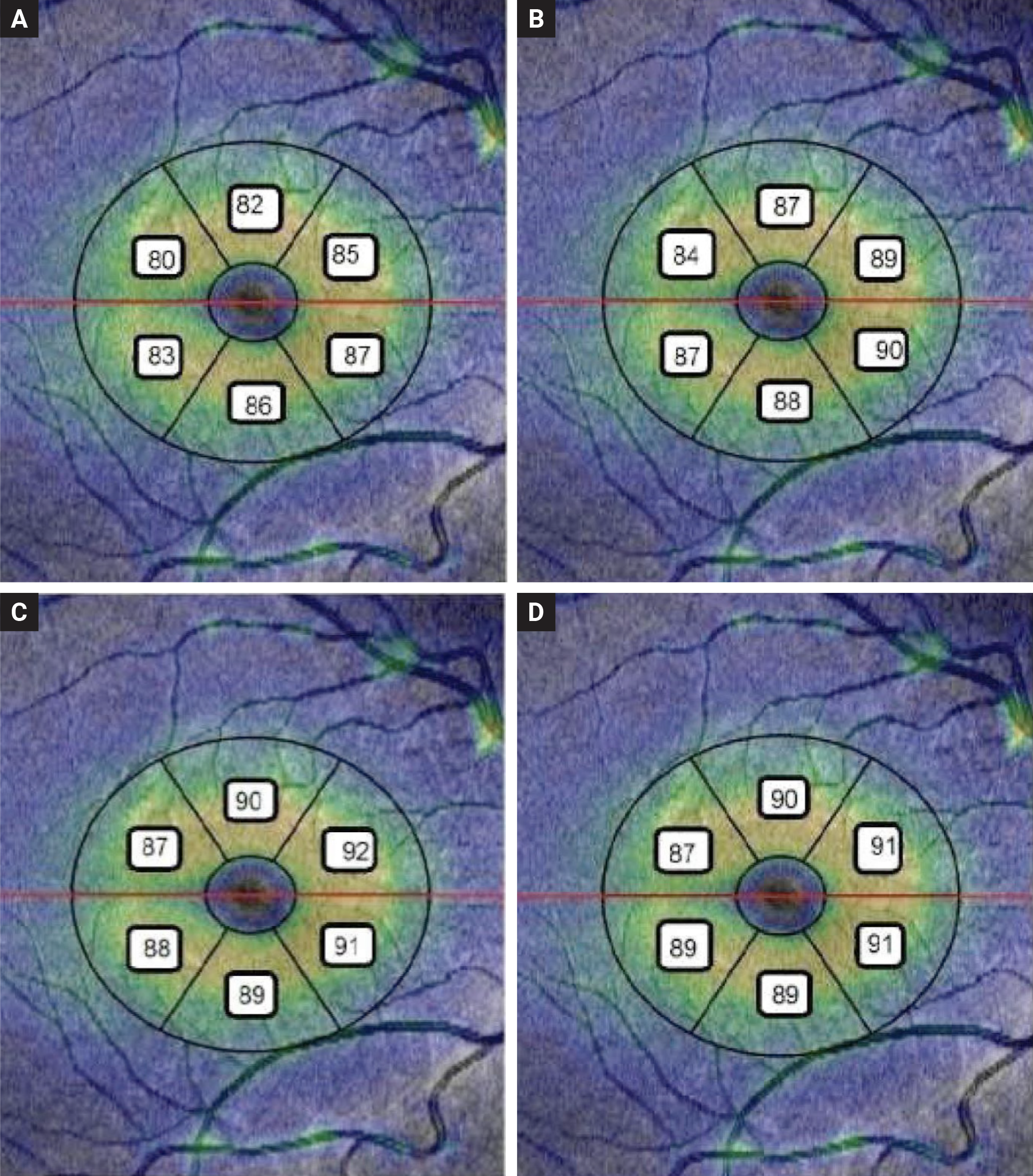
| Children with a history of retinopathy of prematurity (ROP) display unique eye characteristics, such as shorter axial lengths and thicker macula, according to a recent study published in the Journal of Ophthalmology. This image from the study shows thee mean GCL + IPL thickness of six sectors in four studied group: (a) treated ROP; (b) regressed ROP; (c) preterm without ROP; (d) full-term children. Photo: Najjaran M, et al. J Ophthalmol. Sept 30, 2024. Click image to enlarge. |
A recent analysis—published in Journal of Ophthalmology—found that children with a history of retinopathy of prematurity (ROP) exhibited a shorter axial length, steeper cornea and thicker macula, which correlated with lower gestational age.
This retrospective cohort study compared preterm children (aged 4 to 8 years) with or without retinopathy of prematurity and assessed their connections with age and gender-matched full-term children. Researchers measured the following: best corrected visual acuity (BCVA), spherical equivalent (SE) refraction, macular and choroidal thickness, and biometric parameters.
Study participants included children with a history of preterm birth, including ROP who received intravitreal bevacizumab treatment; children with a history of retinopathy of prematurity that regressed without treatment; and children with no history of ROP. Age- and gender-matched full-term children acted as the control group.
A total of 120 eyes of 120 children—30 children in each group—were involved in this analysis. Study authors observed no significant difference in BCVA, SE and subjective cylinder between groups. They found that axial length was significantly shorter and the cornea was steeper among both ROP cohorts when compared to children in the other groups.
Additionally, data showed that the central macular thickness was significantly thicker in the treated, regressed retinopathy of prematurity and preterm groups versus their full-term counterparts. A negative correlation between gestational age and macular thickness was seen in both the treated and regressed ROP patient groups.
While summarizing their findings in their paper on the work, the study authors noted that shorter axial length and steeper cornea as well as a negative correlation between gestational age and macular thickness were reported among treated and regressed ROP children; however, no significant difference in SE or refractive cylinder was observed between the groups.
“This study documents the change in refractive error as well as macular abnormalities, and biometric differences in children with ROP with or without prior treatment and provides valuable information for healthcare providers and funders planning screening and follow-up of these children, particularly during preschool years and during the time period where these children are particularly prone to developing amblyopia,” they wrote.
“Future studies with larger cohorts are required to directly investigate the effect of various retinopathy of prematurity treatments on biometric and OCT characteristics of children and their final impact on visual acuity.”
| Click here for journal source. |
Najjaran M, Zarei-Ghanavati S, Ostadimoghaddam H, et al. Ocular Biometric and Optical Coherence Tomography Parameters in Former Preterm Children: A Cohort Study. J Ophthalmol. September 30, 2024 [Epub ahead of print]. |