 |
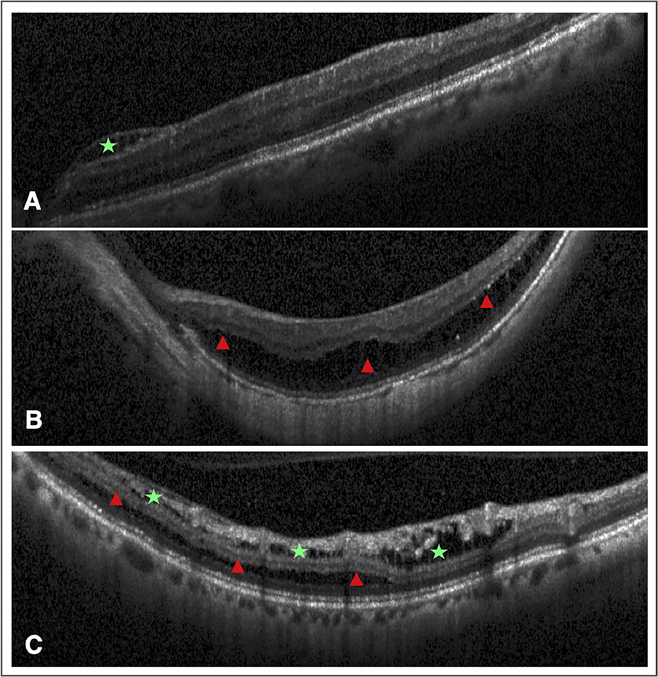
Research shows a significant prevalence of macular retinoschisis in elderly patients with high myopia, identifying critical risk factors and the need for vigilance to preserve visual outcomes. This image from the study illustrates two MRS types: (A) inner retinoschisis, affecting layers from the retinal nerve fiber to the inner nuclear layer (green star), and (B) outer retinoschisis, involving layers from the outer plexiform layer to the retinal pigment epithelium (red triangle). Cases with both types concurrently in the same eye are noted (C). Photo: Pan Z, et al. Am J Ophthalmol. 2024. Click image to enlarge. |
A recent analysis conducted in China found that macular retinoschisis (MRS) affects 27.9% of elderly individuals of Asian descent with high myopia. The study also showed that MRS located in the foveal region or involving the outer retina was linked to a significant decline in best corrected visual acuity (BCVA), highlighting the need for greater clinical attention.
These findings, recently published in American Journal of Ophthalmology, offer new insights into the underlying mechanisms of macular retinoschisis and the condition’s impact on vision prognosis in highly myopic individuals.
This population-based, cross-sectional study examined the prevalence, characteristics and risk factors of macular retinoschisis in highly myopic eyes, as well as their morphological features in a Chinese population. In this analysis, high myopia was defined as at least -6D on refraction or an axial length ≥26.0mm.
Among 213 highly myopic eyes from 129 individuals, MRS was observed in 48 eyes and 36 participants, with a prevalence of 22.5% per eye and 27.9% per subject. Risk factors for macular retinoschisis were identified as higher intraocular pressure, thinner subfoveal choroidal thickness, a wider Gamma zone, glaucoma and the presence of epiretinal membrane.
The study authors reported that development of foveoschisis was more likely among eyes with advanced myopia and a wider gamma zone. Data also showed that eyes with glaucoma had a higher prevalence of outer retinal vs. inner retinal macular retinoschisis; however, the researchers noted that this difference was not statistically significant.
Additionally, the analysis revealed that macular retinoschisis in the foveal region or involving the outer retina was significantly associated with worse best-corrected visual acuity compared to macular retinoschisis that was perifoveally located or affected other regions.
In an elderly Chinese population with high myopia, the study identified a 27.9% prevalence of macular retinoschisis per subject, the study authors summarized in their recent AJO paper. After adjusting for age, myopic refractive error and intraocular pressure, this analysis confirmed a correlation between MRS and the presence of glaucoma and epiretinal membrane.
“Notably, this study is the first to identify an association between the width of the gamma zone and subfoveal choroidal thickness with macular retinoschisis,” the research team noted in their paper. “This suggests that mechanical strains, such as inward and outward tractional forces and sagittal strain within the retinal layers, may be closely linked to the occurrence of macular retinoschisis.
“The findings from current study underscore the importance of detailed anatomical assessments in high myopia patients, enhancing our comprehension of the consequences of MRS and potentially providing a basis for MRS risk assessment and vision prognosis in myopic patients,” they concluded.
| Click here for journal source. |
Pan Z, Huang Y, Li Z, et al. Prevalence, Features and Risk Factors of Macular Retinoschisis in High Myopic Population: the Beijing Eye Study 2011. Am J Ophthalmol. 2024. [Epub ahead of print October 8, 2024]. |