A new analysis highlights the potential role of progressive choriocapillaris changes as a biomarker for age-related macular degeneration (AMD) progression. The data, recently published in the journal Investigative Ophthalmology & Visual Science, showed that widespread choriocapillaris alterations may precede progression to geographic atrophy and more central choriocapillaris loss could provide an ischemic stimulus for wet AMD.
While swept-source OCT angiography (OCT-A) has significantly improved our understanding of AMD pathogenesis, there remains a lack of longitudinal OCT-A data examining choriocapillaris with prolonged follow-up. Therefore, a team of researchers from Harvard, Stanford and Tulane initiated a retrospective observational study to explore the association between inner choroid flow deficit percentage (IC-FD%), various OCT biomarkers and AMD progression in patients with dry, non-advanced disease over a mean follow-up period of 36 months. “A greater IC-FD% correlates with AMD severity, is associated with greater risk of drusen formation or enlargement, and may predict AMD progression ultimately,” they wrote in their IOVS article.
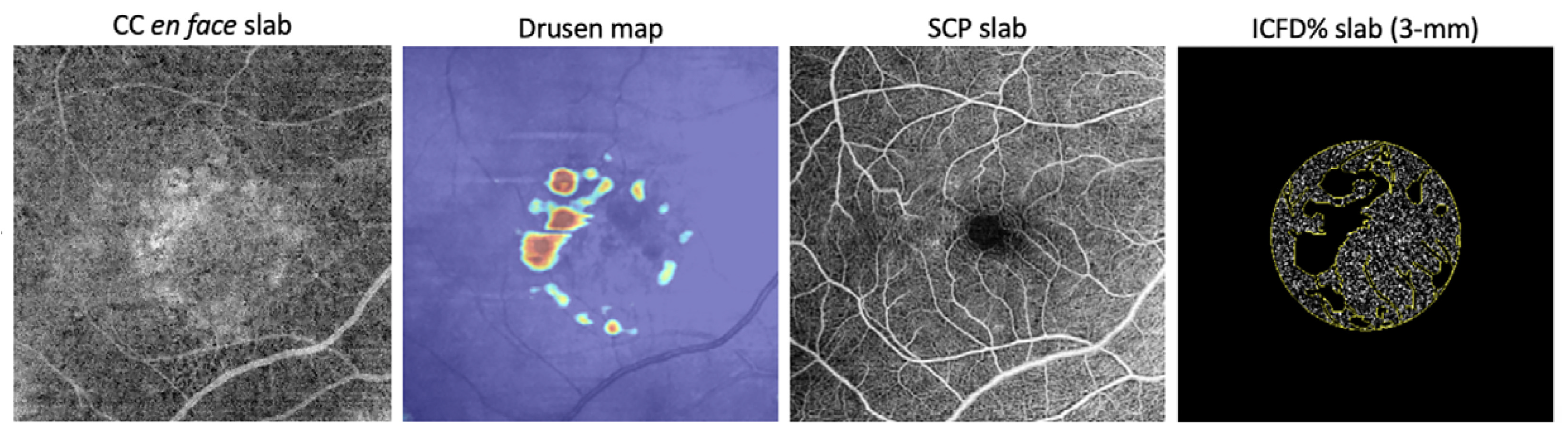 |
| The researchers began with conventional OCT-A imaging of the choriocapillaris, then isolated drusen and the superficial capillary plexus to highlight the structures that might exert a shadowing effect. The IC-FD% was then measured in the central 3mm and 6mm regions. Finally, the team correlated IC-FD% with AMD stage. Photo: Romano F, et al. Invest Ophthalmol Vis Sci. July 11, 2024. Click image to enlarge. |
The analysis included 42 patients (64 eyes) with early or intermediate age-related macular degeneration at baseline. Study participants had two or more consecutive OCT-A covering a period of at least 18 months.
Investigators reviewed demographics, visual acuity and AMD staging based on Beckman classification. They analyzed OCT for hyperreflective foci, subretinal drusenoid deposits, hyporeflective drusen cores and subfoveal choroidal thickness.
During follow-up, data showed that four eyes (31%) with early AMD progressed to intermediate AMD and 30 eyes (59%) with intermediate disease developed late age-related macular degeneration (19 geographic atrophy; 11 wet AMD).
The researchers observed an association with baseline hyporeflective drusen core and geographic atrophy development. Greater inner choroid flow deficit percentage was associated with wet AMD. Additionally, the analysis revealed that faster subfoveal choroidal thickness reduction and inner choroid flow deficit percentage (6mm) increase was correlated with GA, while IC-FD% (3mm) increase was linked with development of wet AMD.
Of note, greater inner choroid flow deficit percentage increases in the 3mm and 6mm were better predictive of wet AMD and geographic atrophy development, respectively, according to the study authors’ Investigative Ophthalmology & Visual Science report.
The researchers noted that their study provides the longest longitudinal assessment of choriocapillaris alterations’ impact on AMD progression using OCT-A. “The conversion to wet AMD was strongly associated with greater inner choroid flow deficit percentage in the central 3mm at baseline and over time, whereas the development of complete RPE and outer retinal atrophy seemed to be predicted better by the identification of hyporeflective drusen core at baseline and a higher rate of inner choroid flow deficit percentage increase across the macular area.”
While the findings may not be definitive due to potential underpowering, the research offers further insight into the pathophysiology of AMD and supports the connection between progressive choriocapillaris insufficiency and increased disease severity, the investigators argued.
“This work emphasizes the importance of choriocapillaris alterations as potential biomarker for risk stratification and monitoring in future clinical trials, paving the way for more targeted and effective interventions in AMD,” the research team concluded in their Investigative Ophthalmology & Visual Science paper.
Romano F, Ding X, Yuan M, et al. Progressive Choriocapillaris Changes on Optical Coherence Tomography Angiography Correlate With Stage Progression in AMD. Invest Ophthalmol Vis Sci. July 11, 2024 [Epub ahead of print]. |