 |
|
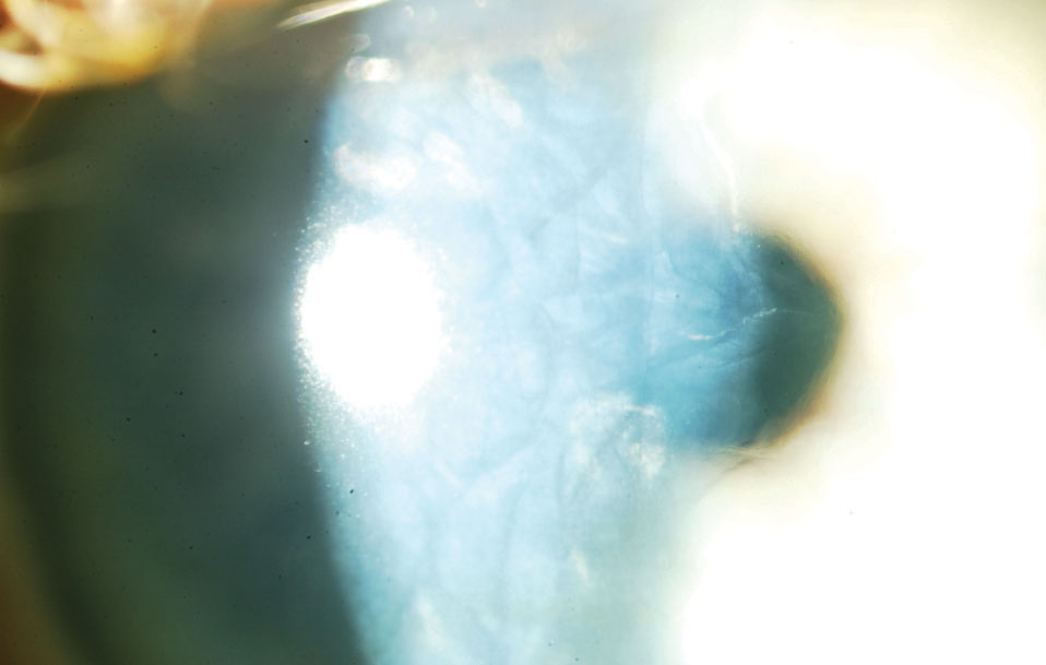
Data from a recent analysis shows significant changes in high-order aberrations in patients with Fuchs endothelial dystrophy who have subclinical corneal edema, underscoring the importance of tomographic analyses in early stages of FECD. Photo: Paul M. Karpecki, OD. Click image to enlarge. |
A new analysis showed significant changes in high-order aberrations (HOAs) among Fuchs’ endothelial corneal dystrophy (FECD) patients with subclinical corneal edema. This data, recently published in the journal Cornea, highlight how tomographic analysis can support visual impairment assessment, disease progression and decision-making for early endothelial keratoplasty in these individuals.
This retrospective, single-center case series, which analyzed whether tomographic changes in FECD impact the HOA in the early period of the disease, included a total of 78 eyes of 47 patients with Fuchs endothelial dystrophy with slit-lamp biomicroscopically visible guttae, but no visible corneal edema. These patients were assigned to group 0 (no subclinical corneal edema) or 1 (subclinical corneal edema).
Group 1 was comprised of 50 eyes of 33 patients with a mean age of 70.9 years and group 0 included 28 eyes from 18 patients with a mean age of 63.9 years. In group 0 and 1, 13 eyes and 34 eyes were pseudophakic, respectively.
Sixty-six healthy eyes of 38 patients were also included in the analysis as the control group.
Of these, 58 eyes were phakic and eight were pseudophakic. Study authors calculated mean values and standard deviations for the root mean square (RMS), coma, trefoil and spherical aberrations of the cornea, the anterior surface and the posterior surface.
Data demonstrated statistically significant differences in the RMS HOA, coma and spherical aberrations of the posterior surface in group 1 eyes when compared to those in group 0. No statistically significant differences in high-order aberrations were observed between the control group and eyes without subclinical corneal edema (Group 0).
These findings showed statistically significant increases in high-order aberrations, particularly of the posterior corneal surface, in patients affected by Fuchs endothelial dystrophy with subclinical corneal edema, according to the study authors who noted, “Our study adds to the current literature by providing evidence of significant changes in high-order aberrations in patients with FECD with subclinical corneal edema compared with those without.
“Early tomographic analysis can, therefore, aid in detecting subclinical corneal edema with varying magnitude of high-order aberrations compared with patients with Fuchs endothelial dystrophy without subclinical edema or healthy eyes, which in turn can help characterize the patients’ visual impairment and decision-making for early endothelial keratoplasty,” they concluded in their recent Cornea paper.
Blöck L, Son HS, Köppe MK, et al. Corneal High-Order Aberrations in Fuchs Endothelial Corneal Dystrophy and Subclinical Corneal Edema. Cornea. July 30, 2024 [Epub ahead of print]. |