 |
|
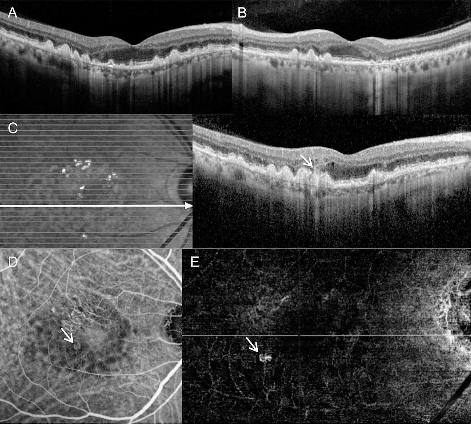
The authors of a new study performed both OCT crosshair and raster scans on the fellow eyes of 75 patients with unilateral nAMD and found that raster scans were more accurate in diagnosing neovascularization in those eyes. A total of 12 cases were missed by OCT crosshair scans alone, and detection was more likely to be missed in patients with type 3 MNV. The authors propose OCT raster scans as a routine component of fellow-eye monitoring. These images from the study show a representative case of a patient diagnosed with type 3 MNV in the left eye. Twelve months after the initial diagnosis, there is no sign of fellow-eye neovascularization on horizontal (A) or vertical (B) crosshair OCT images. However, raster scan images show an intraretinal hyperreflective lesion (C, short arrow) accompanied by adjacent retinal edema and disruption of the RPE inferotemporal to the fovea. Type 3 MNV is observed on indocyanine-green angiography (D, arrow) and OCT-A (E, arrow). The long horizontal arrow on panel C indicates the OCT scanning line. Photo: Kim JH, et al. BMC Ophthalmol. August 21, 2024. Click image to enlarge. |
OCT has become essential to the visualization of the retina, allowing for the magnification of structures in hopes of detecting disease in earlier stages. In cases of unilateral neovascular age-related macular degeneration (AMD), the fellow eye is likely to be affected over time. However, these diagnoses are being missed. In fact, a recent study published in BMC Ophthalmology found that one-sixth of fellow-eye neovascularization cases weren’t initially detected on OCT crosshair scans, but were found with the use of raster scans. This risk was 9.6 times greater in patients with type 3 macular neovascularization (MNV), suggesting raster scans may be beneficial when used routinely.
This study, conducted in Seoul, enrolled 75 treatment-naive patients diagnosed with nAMD and initially treated with three loading injections of ranibizumab or aflibercept. Those who developed fellow-eye neovascularization during the follow-up period (36.6 ±8.5-month) were included (n=75). Among them, the first-involved eye was diagnosed with typical nAMD, polypoidal choroidal vasculopathy (PCV) and type 3 MNV, in 25, 15 and 35 patients, respectively. The mean interval between initial diagnosis and fellow-eye neovascularization was 19.2 ±10.1 months.
Raster scans successfully detected fellow-eye neovascularizations on all patients, whereas 16% of eyes were missed on crosshair scans. Among the 35 fellow-eye neovascularization cases in patients with type 3 MNV, 28.6% were missed by crosshair scans. “We believe that the primary reason for this finding is the characteristic lesion distribution in type 3 MNV,” the authors wrote in the study. “It is well known that type 3 MNV lesions typically develop with foveal sparing. The absence of a deep capillary plexus in the foveal region, which is postulated to be the origin of type 3 MNV, may contribute to the distinctive distribution pattern of the lesions. For prompt diagnosis of type 3 MNV, OCT should be performed directly at the site of the lesion. This is particularly crucial when a lesion develops relatively distant from the fovea.”
OCT crosshair scans, while rapid, may be limited in that they are centered horizontally and vertically on the fovea; whereas raster scans enable a more detailed assessment through multiple horizontal scans of the macula, but require more time and effort to obtain. With this in mind, the study authors say, given the relentless progressive nature of type 3 MNV, early diagnosis is crucial. “Considering the markedly high risk of fellow-eye neovascularization in type 3 MNV, it is imperative to perform raster scans and meticulously examine all acquired images during fellow-eye screening, even if it entails additional time and effort,” they wrote.
Screening frequency is also a consideration. “In this study, fellow-eye neovascularization was not detected on crosshair scans when the interval between fellow-eye examinations was shorter,” said the authors. “It is possible that more frequent screenings may have led to the detection of neovascularization at an earlier stage, thus influencing study outcomes.” They added, to date, there’s no established consensus for the optimal interval of fellow-eye exams in cases of unilateral nAMD.
Lack of monthly follow-up may have contributed to the delay in fellow-eye neovascularization detection, which the authors cite as a possible limitation. They also mention other limitations, including the clinical setting in which treatment data was obtained, as well as the fact that all participants were Korean. Also, they mention that “OCT angiography, which exhibits good sensitivity for detecting neovascularization in its early stages, was not routinely performed for fellow-eye monitoring.”
Despite this, due to the risk of undetected neovascularization on crosshair scans being markedly high for patients whose initially affected eye has type 3 MNV, the authors say routine OCT raster scans are necessary during fellow-eye examinations in all cases of unilateral nAMD, but especially in this group, they concluded.
Kim JH, Kim JW, Kim CG. Importance of optical coherence tomography raster scans in early detection of active fellow-eye neovascularization in unilateral neovascular age-related macular degeneration. BMC Ophthalmol. August 21, 2024. [Epub ahead of print.] |