 |
|
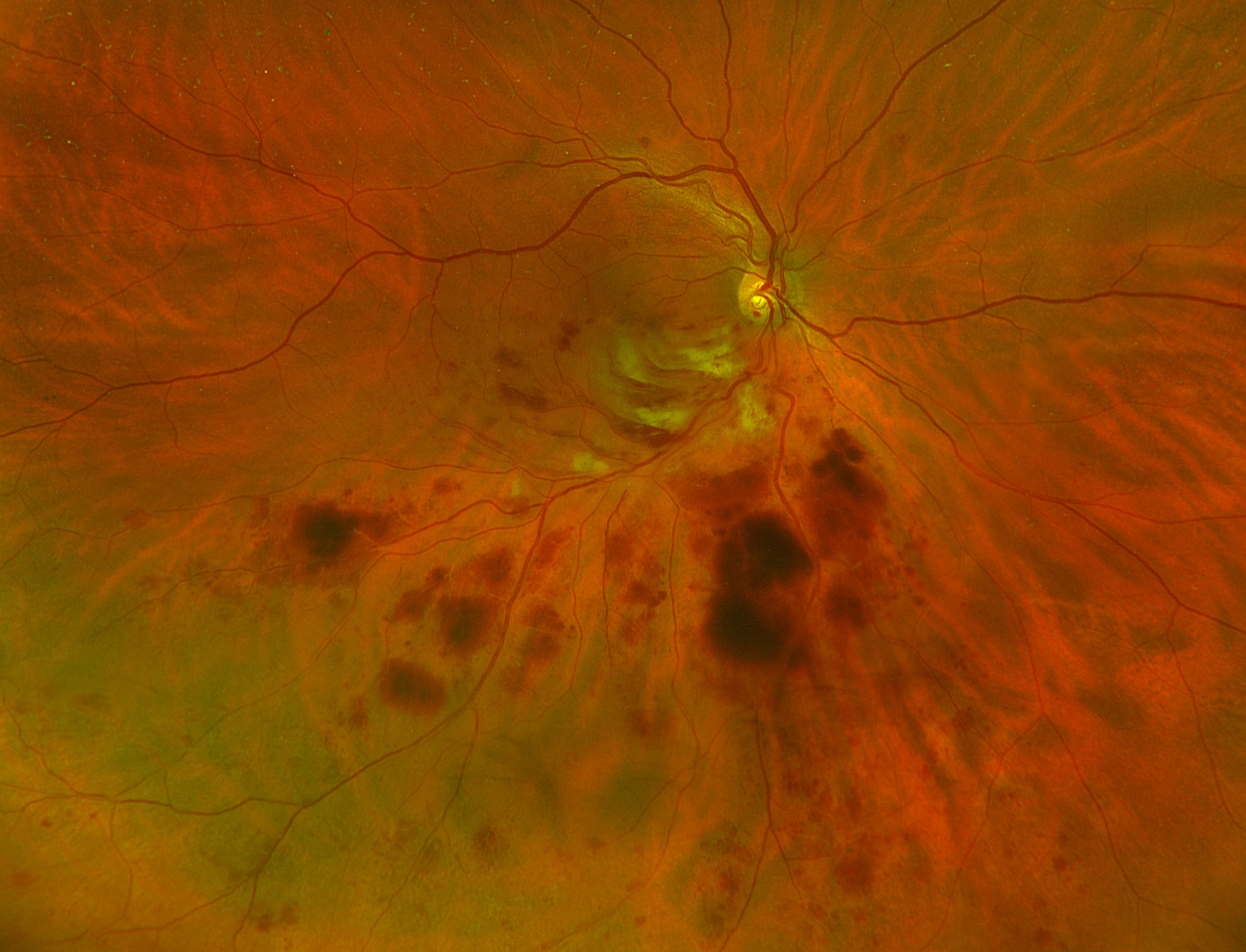
A new analysis showed that structural changes in the choroid among RVO patients receiving intravitreal anti-VEGF therapy could be useful tool during clinical follow-up of these individuals. Photo: Rami Aboumourad, OD. Click image to enlarge. |
A recent analysis suggests that changes in choroidal structure may help clinicians assess retinal vein occlusion (RVO) and predict the efficacy of anti- VEGF therapy. These findings showed that a metric known as choroidal vascularity index (CVI) was lower in patients with RVO compared to controls; however, it increased significantly with anti-VEGF treatment, especially at one month following injection. CVI quantifies the vascular component of the choroid (by measuring vessels’ luminal area) as a percentage of the entire structure.
“The choroidal thickening observed in retinal vein occlusion appears to result from stromal edema, possibly triggered by elevated intraocular vascular endothelial growth factor levels, rather than direct vascular dilatation,” the study authors reported in the journal Graefe's Archive for Clinical and Experimental Ophthalmology,
This prospective longitudinal study explored how RVO impacts the structures of the posterior segment of the eye and to assess the changes that occur with intravitreal anti-VEGF therapy.
In this analysis, 29 eyes of 29 patients (17 male, 12 female) with retinal vein occlusion were followed for six months. The best corrected visual acuity (BCVA), macular thickness, choroid thickness and CVI obtained by OCT were assessed at baseline and the first, third and sixth months following the first injection. Researchers compared results with fellow (non-affected) eyes as well as age- and sex-matched controls.
Data showed that BCVA increased significantly in the sixth month, more in the first month of injection, according to the study authors, who also reported a significant decrease in central macular thickness, subfoveal choroid thickness, stromal and total area of choroid following injection.
Additionally, there a significant increase in CVI, particularly in the first month after injection. Among eyes with branch RVO, results revealed a significant decrease in the macular thickness of the occlusive areas with treatment; however, no statistically significant change was observed in the non-occlusive macular thickness.
“The increased choroidal thickness in eyes with macular edema associated with retinal vein occlusion was primarily caused by stromal area rather than choroidal lumen area and decreased after anti-VEGF treatment,” the study authors noted in their recent Graefe's Archive for Clinical and Experimental Ophthalmology paper.
“In eyes with RVO, lumen area remains unchanged, stromal area increases, secondary CVI decreases and choroidal thickness increases,” they continued. “Contrary to initial assumptions, the observed choroidal thickening in retinal vein occlusion appears primarily driven by stromal edema, likely triggered by elevated intraocular VEGF levels, rather than direct vascular dilatation.”
Dursun E, Derhem B, Çobanoğlu S, et al. Investigation of choroidal structure changes after intravitreal anti-VEGF therapy for retinal vein occlusion. Graefes Arch Clin Exp Ophthalmol. July 22, 2024 [Epub ahead of print]. |