 |
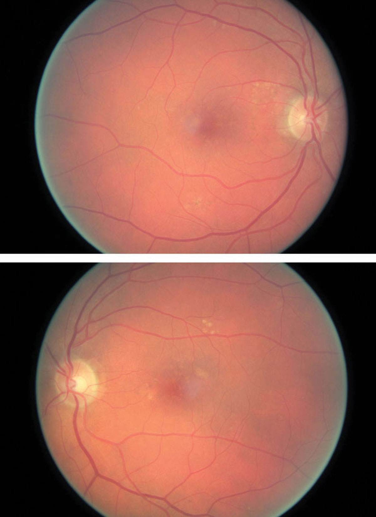
| Peak emission wavelength may play a role in AMD diagnosis. Photo: Amanda Legge, OD. Click to enlarge. |
Fundus autofluorescence (FAF) imaging has become part of routine clinical diagnostics in recent years, but there is limited knowledge on its role in the pathophysiology of retinal disease. In a recent study, researchers investigated the spectral characteristics of FAF in AMD patients and healthy subjects.
FAF spectral characteristics were described by the peak emission wavelength (PEW) of the spectra. PEW was derived from the ratio of FAF recordings in two spectral channels at 500nm to 560nm and 560nm to 720nm by fluorescence lifetime imaging ophthalmoscopy. PEW measurements were done in 44 younger healthy participants with a mean age of 24, 18 elderly healthy subjects with a mean age of 67.5 and 63 patients with a mean age of 74 with AMD.
No significant difference was found between PEW in young and elderly controls. However, PEW was significantly shorter in AMD patients. At follow-up, PEW decreased significantly in the inner ring of the grid. Patients, showing progression to atrophic AMD during follow-up, had significantly shorter PEW at baseline than non-progressing patients.
“Our findings indicate a slight hypsochromic emission shift with aging and significantly shorter emission wavelengths in AMD vs. controls,” the study authors concluded. “Patients with progression to outer retinal atrophy (ORA) or complete outer retinal atrophy (cRORA) at follow-up show shorter PEW in the baseline measurement already.”
Similar to a previous study, the rate of disease progression was higher in the patients showing subretinal drusenoid deposits, and these patients had insignificantly lower PWE.
Another study determined lipofuscin as the dominant retinal fluorophore but also found a minor fluorophore with peak emission at 520nm to 540nm, which could possibly be related to flavin adenine dinucleotide, the authors noted. This fluorophore was seen most clearly where lipofuscin was low in the macula, which was found in this current study of shorter PEW in the macula of healthy subjects.
“When lacking lipofuscin fluorescence, FAF is dominated by contributions of Bruch’s membrane and remaining basal linear and laminar deposits, eventually also from the retina and deeper layers such as the choroid and sclera. For this reason, we excluded patients with ORA and cRORA from the comparison with healthy controls, as well as from the analysis of spectral changes of FAF in the follow-up of AMD,” the authors noted.
“In conclusion, PEW might be a diagnostic marker in AMD,” the authors said. “It was not only shorter in the patients than in the controls, but also shorter in patients who later progressed to ORA or cRORA than in those who did not. Thus, short PEW might be an indicator for AMD progression.”
Schultz R, Schwanengel L, Meller D, Hammer M. Spectral fundus autofluorescence peak emission wavelength in aging and AMD. Acta Ophthalmol. December 1, 2021. [Epub ahead of print.] |