 |
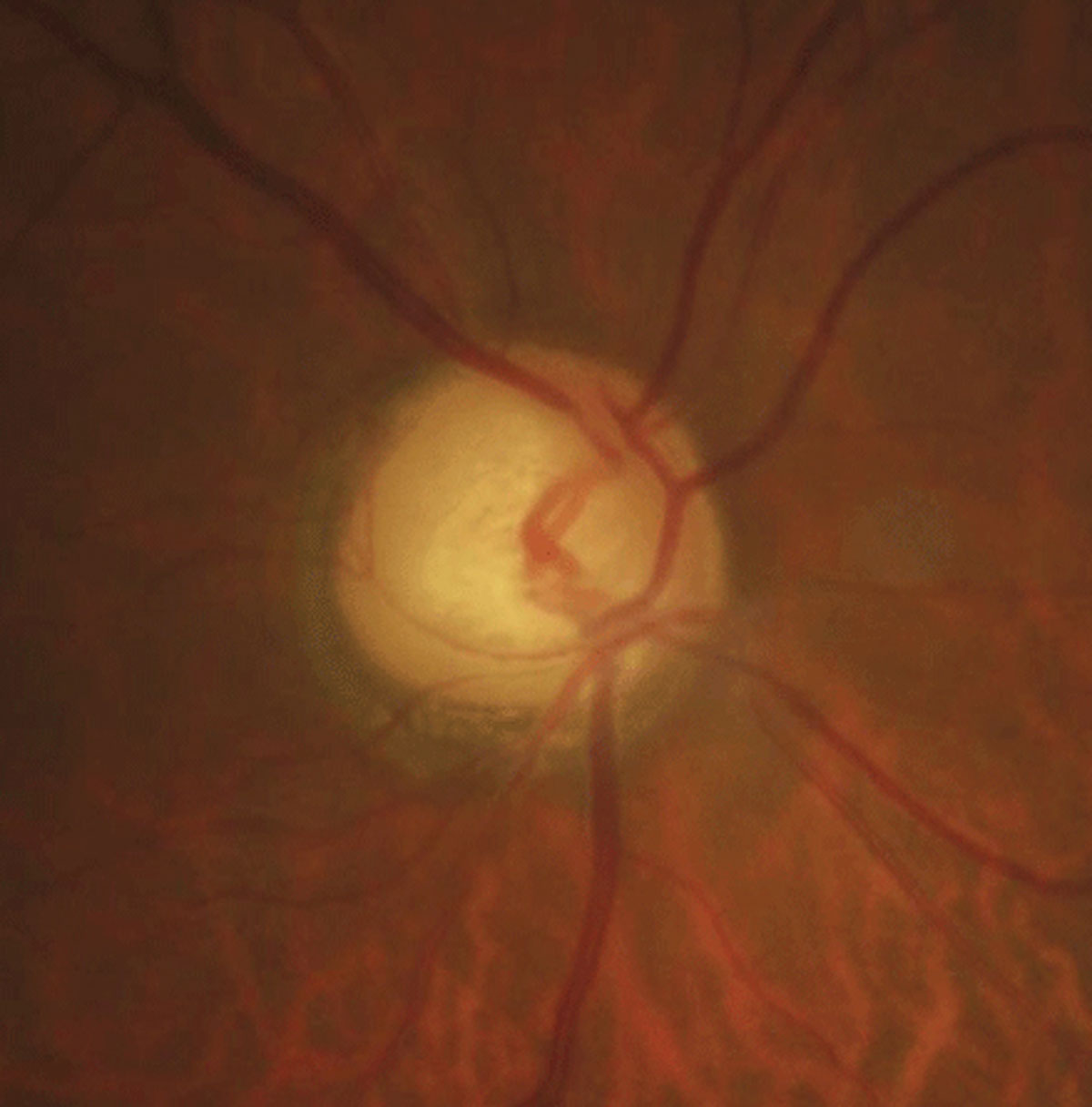
| Chromatic pupillometry helps identify abnormal light responses in glaucoma. Photo: Justin Cole, OD, and Jarett Mazzarella, OD. Click to enlarge. |
Optometric research is continually looking for tangible ways to detect and treat glaucoma sooner, as the condition is often diagnosed at a later stage resulting in more negative outcomes and less effective options for intervention. Chromatic pupillometry has become an increasingly popular method of analyzing retinal and optic nerve health in recent years by measuring pupillary responses to colored light stimuli. Now, research is showing that this technique may be able to accurately reveal functional loss in glaucoma even in the earliest stages of the disease, giving it potential as a reliable, inexpensive screening tool.
By evaluating the performance of retinal photoreceptors (rods, cones and intrinsically photosensitive retinal ganglion cells (ipRGCs) with a handheld chromatic pupillometer (HCP), loss or dysfunction of ipRGC may be detected, which leads to an abnormal pupillary response and signifies the presence of glaucoma in a patient. Researchers put this technique to the test by ranking its ability to identify and classify glaucoma, as well as by comparing it to other structural and functional investigational tools for the disease including optical coherence tomography (OCT) and Humphrey Visual Field (HVF).
The clinic-based, prospective study included 149 glaucoma patients and 173 healthy controls. Using a custom-built HCP, the team evaluated patients’ changes in pupil size in response to nine seconds of exponentially increasing blue (469nm) and red (640nm) light stimuli during a one-minute pupillometric self-examination. Dynamic pupillary responses were all observed without dark adaptation, and the tests were performed in a dimly lit room.
The findings revealed that pupillary light responses were altered in glaucoma patients compared with controls and that this technique can effectively detect the distinction. After researchers performed receiver operating characteristic (ROC) curve analyses, they found that HCP, overall, showed a similar level of accuracy as both HVF and OCT in its glaucoma detection and classification capabilities.
“For glaucoma classification, HCP yielded an area under the ROC curve (AUC) of 0.94, sensitivity of 87.9% and specificity of 88.4%,” the researchers wrote in their paper on the study. “The classification performance of HCP in early-moderate glaucoma (visual field mean deviation >-12dB) was similar to HVF (AUC=0.91) and reduced compared with OCT (AUC=0.97). For severe glaucoma (VFMD ≤-12dB), HCP had an excellent classification performance (AUC=0.98), similar to HVF and OCT.”
The researchers noted that this screening method can even be done reliably on patients with mild to moderate cataracts or refractive errors and could be a more time- and cost-effective approach for detecting abnormal pupillary responses in patients with or suspected to have glaucoma.
“In this proof-of-concept study, we show that a low-cost handheld pupillometer delivering short-duration light stimulations, requiring minimal fixation aptitudes and no dark-adaptation, can highlight functional loss in eyes with glaucoma,” the research team concluded.
Najjar RP, Rukmini AV, Finkelstein MT, et al. Handheld chromatic pupillometry can accurately and rapidly reveal functional loss in glaucoma. Br J Ophthalmol. November 6, 2021. [Epub ahead of print]. |