 |
|
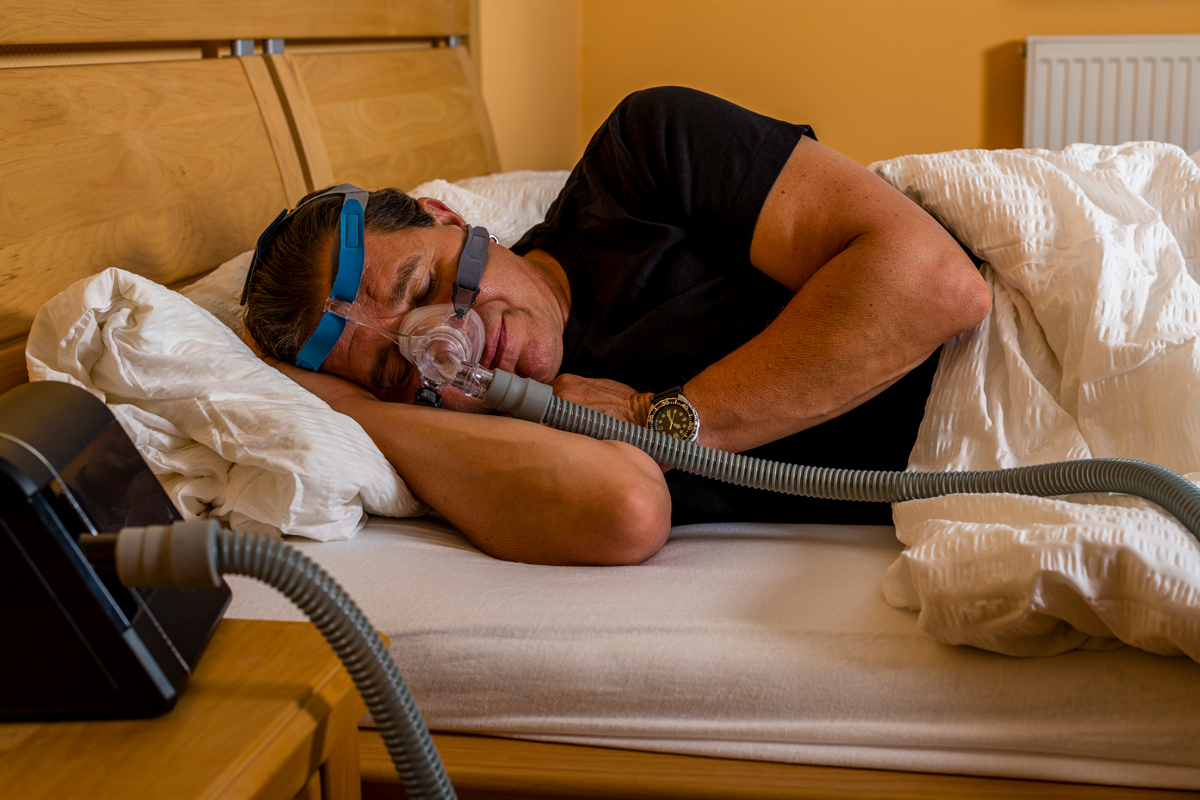
Findings suggests a connection between peripapillary vascular density and disease severity among patients with obstructive sleep apnea-hypopnea syndrome. Photo: Getty Images. Click image to enlarge. |
A recently published study found that patients with obstructive sleep apnea-hypopnea syndrome (OSAS) showed reduced peripapillary vascular density. Based on these results, the researchers propose that measuring optic nerve head vascular density using OCT angiography (OCT-A) could be a useful method for diagnosing and monitoring the disease severity among these patients.
In this meta-analysis, researchers sought to evaluate retinal microvasculature alterations in patients with OSAS, offering further insight to help clinicians better understand and assess this population of patients.
A literature search of several electronic databases initially identified 134 studies. Of those, six eligible articles, comprising 479 eyes (333 in the OSAS group and 146 in the control group) were included in the meta-analysis.
Pooled results showed that radial peripapillary capillary (RPC) whole en face vessel density (VD) was significantly decreased in the mild-to-moderate OSAS patient group when compared to the control group. “For RPC peripapillary VD, eyes in mild-to-moderate OSAS showed a trending decrease compared to the controls, and there was a remarkable difference between eyes with severe OSAS and the controls,” the study authors reported in Journal of Ophthalmology.
The investigators also analyzed the difference between the severe sleep apnea cohort and controls. They found that radial peripapillary capillary inside disc vessel density was decreased among eyes with severe OSAS vs. patients in the control group.
“To date, this is the first meta-analysis to calculate changes in retinal microvasculature density measured by OCT-A in patients with obstructive sleep apnea-hypopnea syndrome and healthy controls,” wrote the researchers.
“Our current results demonstrated that peripapillary vascular density was attenuated in patients with OSAS,” they noted in their recent Journal of Ophthalmology paper. “Moreover, on the basis of these findings and the noninvasive nature of OCT-A imaging facility, we suggest that the measurement of optic nerve head vascular density by OCT-A may presumably serve as a potential biomarker to objectively monitor and diagnose obstructive sleep apnea-hypopnea syndrome in the future.”
Ji K, Yang Yang Y, Zhang Q, et al. Meta-Analysis: Characteristics of retinal vasculature in obstructive sleep apnea syndrome humans. J Ophthalmol. July 16, 2024 [Epub ahead of print]. |


