 |
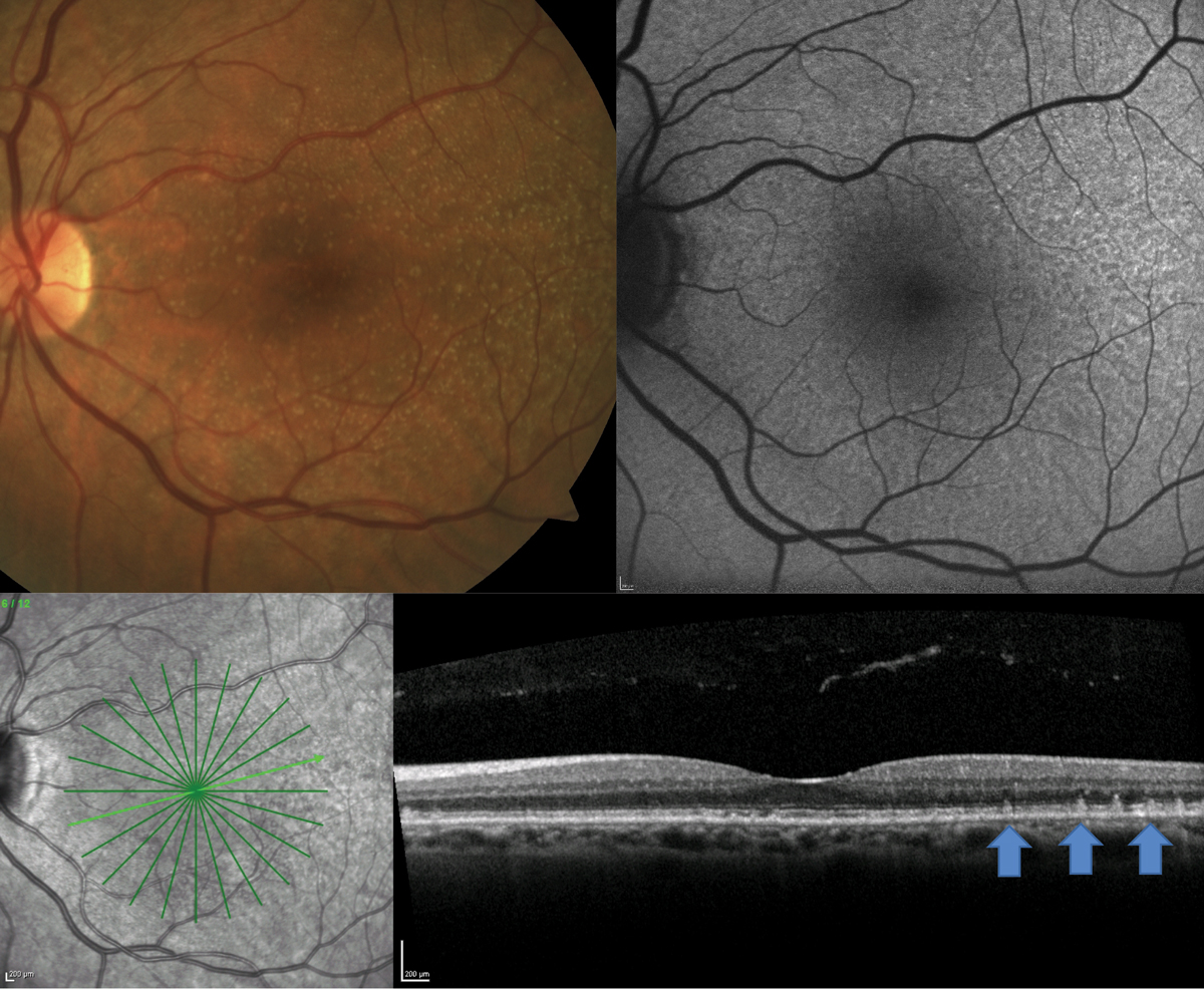
| A patient with clinically visible small- to medium-sized drusen shows presence of RPD on FAF and OCT. Photo: Jessica Haynes, OD. Click image to enlarge. |
Beyond the importance of specific biomarkers in predicting complete retinal pigment epithelium (RPE) and outer retina atrophy growth, many believe that recognizing unique OCT phenotypes in intermediate AMD would be essential to develop a simplified risk assessment for such outcomes. Researchers in Italy evaluated the impact of OCT phenotypes preceding atrophy related to AMD on the progression of atrophic lesions. The team identified that non-neovascular splitting in the region of RPE, basal lamina and Bruch’s membrane represented the main OCT phenotype involved in the progression of retinal atrophy.
This observational retrospective cohort study, published in Retina, included a total of 70 eyes of 60 consecutive patients with intermediate AMD with a minimum follow-up of 24 months. The atrophy was quantified using fundus autofluorescence, also considering the directionality of atrophy as centrifugal and centripetal progression rates. The phenotype organization was based on the elementary AMD lesions observed on OCT B-scan, including drusen, reticular pseudodrusen/subretinal drusenoid deposits and pigment epithelium detachment (PED), as well as the presence of an RPE/basal lamina/Bruch’s membrane splitting in the absence of neovascular signal using OCT-A.
The best-fit model for GA (odds ratio; OR: 1.81) revealed that the main baseline predictor was the presence of splitting at the junctions of RPE, basal lamina and Bruch's membrane. This phenotype was associated with a faster evolution with a more significant expansion of the atrophic area compared to other phenotypic lesions.
Large drusen at baseline also were prognostic, this time appearing protective for the GA area lesion expansion over time (OR: 0.52) when considered with other confounders. The presence of large drusen was accompanied by a different pattern of GA growth with a greater foveal sparing, probably due to the tendency of spreading around the fovea first, configuring a horseshoe-like pattern. Interestingly, their results indicated that the presence of large drusen resulted in a larger area of foveal sparing and a slower rate of progression.
“By modeling intermediate AMD elementary lesions with a potentiality to evolve into atrophy, we have obtained a simplified risk stratification where the presence of a pseudo double layer sign represented a crucial predictor among others considered,” the authors wrote in their paper. “The recognition of this phenotypic lesion through OCT can delineate a high-risk phenotype characterized by a rapid spreading of atrophy and larger lesions compared with eyes without a double layer sign.”
Fragiotta S, Dysli C, Parravano M, et al. Phenotypic characterization of predictors for development and progression of geographic atrophy using optical coherence tomography. Retina. March 11, 2024. [Epub ahead of print]. |