 |
|
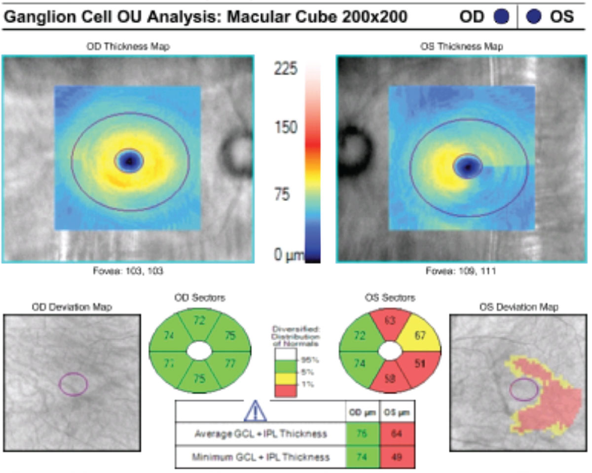
Macular ganglion cell analysis can be a screening tool that can discern glaucoma patients from glaucoma suspects. Photo: Bisant A. Labib, OD. Click image to enlarge. |
The concept of macular ganglion cell analysis for glaucoma diagnosis is gaining favor relative to the more established circumpapillary retinal nerve fiber layer (cRNFL) assessment in determining structural deficits in perimetric glaucoma or as a tool to diagnose pre-perimetric glaucoma. The use of ganglion cell layer (GCL) and inner plexiform layer (IPL) measurements in the latest ganglion cell analysis algorithm aims to diminish the effect of nerve fiber layer measurements, wherein the distribution in the macula is highly dependent on individual anatomy. In a new study, researchers in the Philippines determined the validity of GCL + IPL in the evaluation of glaucoma across different stages using the area under the curve (AUC) analysis in comparison to cRNFL. They found that, compared to cRNFL, macular ganglion analysis may be more beneficial in glaucoma screening and detecting progression from glaucoma suspect to mild glaucoma.
The charts of 260 adult glaucoma suspects and glaucoma patients having macular ganglion cell analysis, OCT of the cRNFL and automated visual field were reviewed. GCL + IPL thickness (average, minimum and sectoral) and sectoral cRNFL thickness were obtained. A total of 122 eyes were included in the study and were grouped into glaucoma suspects (n=43), early or mild glaucoma (n=40) and moderate-to-severe glaucoma (n=39). These were patients without retina or inflammatory pathology and without optic nerve pathology other than glaucoma and were patients with refractive errors within +6.00D hyperopia or -6.00D myopia with a cylinder correction within ±3.00D.
Both GCL + IPL and cRNFL thickness parameters showed a significant decline with greater glaucoma severity. In the determination of visual field defects across all glaucoma stages, the highest AUC was obtained by minimum GCL + IPL (AUC=0.859) with a cut-off value at ≤70μm. Average GCL + IPL had the highest AUC (0.835) in detecting progression from glaucoma suspect to mild glaucoma, while the inferior sector of the cRNFL had the highest AUC (0.937) in discerning mild from moderate-to-severe glaucoma.
“Our study compared the GCL + IPL and cRNFL thickness parameters in glaucoma suspects and across mild to moderate-to-severe glaucoma stages and filled in that gap in knowledge,” the authors of the study, which was published in Clinical Ophthalmology, wrote. “Compared to cRNFL, macular ganglion analysis may be more beneficial in glaucoma screening and detecting progression from glaucoma suspect to mild glaucoma.”
San Pedro MJS, Sousan GMN, Yap-Veloso MIR. Correlation of macular ganglion cell layer + inner plexiform layer (GCL + IPL) and circumpapillary retinal nerve fiber layer (cRNFL) thickness in glaucoma suspects and glaucomatous eyes. Cl Ophthalmol. 2024;18:2313-25. |