 |
|
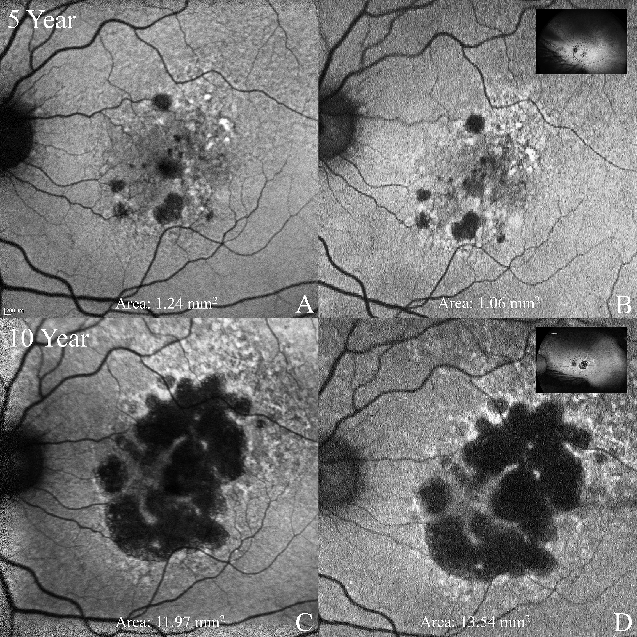
Most features associated with geographic atrophy were comparable between the two tested modalities, except for reticular pseudodrusen, which was less visible on Optos images. These images from the TVST paper show geographic atrophy progression between five-year (A, B) and 10-year (C, D) time points observed on standard field (A, C) and ultra-widefield (B, D) devices. Photo: Froines CP, et al. TVST 2024. Nov. 4, 2024. Click image to enlarge. |
Fundus autofluorescence (FAF) is one of the mainstays of geographic atrophy (GA) monitoring both in the clinic and as a primary endpoint in clinical trials. The standard of care is the standard 30-degree field (Heidelberg Engineering), which uses blue excitation to produce FAF images. Another modality gaining popularity is ultrawide field FAF (Optos), which enables a 200-degree view in a single image and uses green excitation. While GA shows up as hypoautofluorescence on both imaging modalities, the measurement differences between the two devices are unclear. Cross-device comparisons must be carefully considered. To quantify these differences, researchers compared GA measurements and progression over time using the Spectralis and the Optos devices. They confirmed that the devices can’t be used interchangeably and stated that both are suitable for long-term monitoring.
The study included 73 participants with GA from the AREDS2 and Optos PEripheral RetinA (OPERA) studies. Researchers evaluated two time points, five years apart and recorded the GA area in the macular area for both imaging modalities and in the peripheral field for UWF.
For the 102 paired images, the researchers reported a mean baseline GA area of 5.32 mm2 with standard and 4.79 mm2 for UWF FAF, demonstrating a significant mean difference of 0.52 mm2. They also reported that GA progression over five years in 25 eyes showed a median annual growth rate of 1.28 mm2 and 1.34 mm2 for standard and UWF FAF, respectively.
Based on these findings, the researchers concluded in their TVST paper that GA measurements were larger on Heidelberg’s standard field than on Optos’ UWF FAF imaging. They suggested that this discrepancy “may be related to differences in image averaging and use of blue versus green FAF, respectively. Comparable GA progression measurements between modalities suggest that either may be used longitudinally in clinical trials, although not interchangeably.”
| Click here for journal source. |
Froines CP, Saunders TF, Heathcote JA, et al. Comparison of geographic atrophy measurements between blue-light Heidelberg standard field and green-light Optos ultrawide field autofluorescence. TVST 2024. [Epub November 4, 2024]. |


