A recent study sought to determine the differential diagnosis for the presence of foveal hyperreflective vertical lines (FVL) on spectral-domain OCT. The researchers found that the finding was associated with various underlying disorders, though no specific FVL pattern could characterize or distinguish these conditions.
This observational case series study, conducted in Israel, gathered data from the retina unit of a tertiary eye care referral center on 10 eyes of 10 patients with FVL present on their SD-OCT structural images and no history of retinal surgery. According to the researchers, “FVL was defined as any vertical hyperreflective line of more than 40µm at the fovea, at any level from the retinal nerve fiber layer to the pigment epithelium.”
|
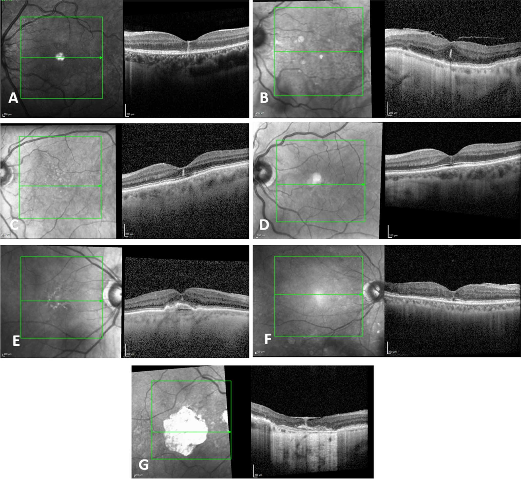
| Researchers observed several different underlying disorders among patients with foveal hyperreflective vertical lines detected on SD-OCT, though no particular line pattern characterized any of the differential diagnoses. This image from the study shows Examples of FVL in different clinical settings: (A) inflammation, (B) mechanical, (C) resorption of fluids, (D) macular telangiectasia, (E) AMD, (F) diabetic retinopathy, (G) scar. Photo: Rein AP, et al. Graefe's Archive for Clinical and Experimental Ophthalmology. October 15, 2024. Click image to enlarge. |
The mean age at presentation was 69 years, and in all patients, FVL were unilateral. The researchers found that the presence of FVL on SD-OCT was associated with tractional disorders of the vitreoretinal interface, accompanying fluid resorption, inflammation, macular telangiectasia, AMD, diabetic retinopathy and scarring. Resorption of fluids (from various causes) was found in the most eyes with FVL (four of 10), while the other underlying conditions were each observed in one eye among the cohort.
“Analysis of those specific cases, in their proper clinical context, enables us to describe various conditions in which FVL may appear,” they continued. The authors then briefly summarized the proposed mechanisms behind the presence of FVL in each differential diagnosis reported in this study.
Despite the unique etiologies of the finding, no particular FVL pattern, “isolated from broader morphological elements or the clinical context,” could characterize any of the underlying conditions or distinguish them from one another, the researchers noted in their paper.
While this study helped identify various ophthalmic disorders associated with FVL, it has several limitations, such as its retrospective nature and small sample size. Therefore, the researchers suggested that future studies be conducted with a larger cohort to evaluate more etiologies for FVL.
“Recognition of this pattern and formulation of an appropriate differential diagnosis is of interest for correctly diagnosing and treating patients whose structural OCT harbors this overlooked finding,” they concluded.
| Click here for journal source. |
Rein AP, Totah H, Brosh K, Zadok D, Hanhart J. Foveal hyper‑reflective vertical lines detected by optical coherence tomography: imaging features, literature review and differential diagnoses. Graefe's Arch Clin Experimental Ophthalmol. October 15, 2024. [Epub ahead of print]. |


