 |
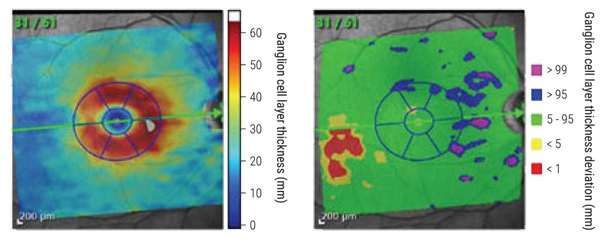
Factors such as a patient's age, sex and axial length had an influence on GCL thickness in patients in this study. Photo: James Fanelli, OD. Click image to enlarge. |
It’s known that different factors can affect ganglion cell layer (GCL) measurements with OCT, but not many studies have examined these in-depth. In a recent study, researchers examined the effect of age, sex and axial length on normal retinal GCL thickness and volume with OCT in a healthy population without any neurological diseases.
One hundred and sixteen eyes of 60 participants between the ages of 12 and 76 (55% female) were examined.
GCL volume was greater in males compared with females after adjusting for age and axial length. GCL volume declined with age but not after adjusting for sex and axial length, and it declined with longer axial length after adjusting for age and sex.
The authors suggested thinning of the GCL layer with increasing axial length can be explained by the distribution of the nearly fixed number of ganglion cells in the retina over a larger surface area in the case of a longer bulb.
GCL thickness differed between males and females in the inner retinal ring but not in the outer ring. “As the ganglion cell develops from the brain, changes in thickness with both age and sex may indicate a possible association between the GCL and brain volume,” the authors explained in their paper on the study. “The effect of sex hormones during the fetal stage or in early infancy may be implicated, as these hormones affect the neural development as well as the neuronal complexity and axonal and process formation in the brain.”
“The GCL thickness difference we found in the various anatomic sectors of the inner ring indicates that the vertical sectors (superior and inferior) are the thickest followed by the nasal and then the temporal; this may reflect the migration pattern of ganglion cells at the macula during development, where the cells adopt a more horizontal migration direction than vertical.”
Lastly, although the OCT was correct in most of the measurements, it should be noted that 25% of the measurements had to be manually adjusted after automated segmentation by OCT.
“Based on our results, it is recommended that future studies examining GCL thickness or volume should take age, sex as well as axial length into consideration, as well as the possible need for manual adjustment of automated OCT measurements,” the authors concluded.
Al-Hawasi A, Lagali N. Retinal ganglion cell layer thickness and volume measured by OCT changes with age, sex and axial length in a healthy population. BMC Ophthalmol. June 24, 2022. [Epub ahead of print]. |