 |
|
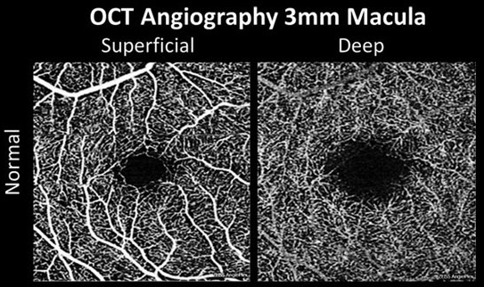
The study found significant differences in FAZ and vessel density-related metrics between white and non-white participants. This difference persisted despite accounting for the socioeconomic status of patients using a well-established index. Photo: Carolyn Majcher, OD. Click image to enlarge. |
The interplay between patient demographics and disease risk profile is complex, as both anatomical and socioeconomic factors influence systemic and ocular health. Researchers from San Francisco studied the impact of racial/ethnic status on the retinal vasculature of diabetes patients using an urban population in a new paper published in Ophthalmic Epidemiology. A total of 2,026 patients were assessed for this study (217 non-diabetic and 1809 patients with diabetes). Then, researchers recorded the different race and ethnic backgrounds of their sample population. They broke down their pool of subjects into five groups: Hispanic (42.2%), Non-Hispanic Asian (24.9%), Non-Hispanic Black (6.8%), Non-Hispanic White (9.7%) and Other (16.3%). Non-Hispanic White patients were used as the reference group for this study.
Using OCT angiography scans to analyze the vascular characteristics of each patient, researchers were able to determine which racial and ethnic groups had more or less significant retinal microvasculature differences. In both the non-diabetic and diabetic groups, Hispanic, Asian and Black patients had significantly greater foveal avascular zone areas and perimeters when compared to white patients, even when controlling for socioeconomic status. Additionally, lower deep capillary plexus vessel density and vessel length density were significantly associated with Hispanic, Asian and Black patients.
This study did have its limitations. Researchers allowed their subjects to self-report this racial and ethnic identities, which they believe this could have led to some bias among patients. Also, each patient was categorized by race and ethnicity, which does not fully constitute biologic or genetic patterns, and each category of race and ethnicity was not evenly distributed in this study. Furthermore, the patient population consisted of non-diabetic retinopathy and mild diabetic retinopathy cases, so the researchers were not able to assess the differences among patients with advanced stages of diabetic retinopathy.
“Our findings underscore the importance of early detection and emphasize the need to develop appropriate screening guidelines, keeping in mind the variations that may exist in our diverse populations,” stated the researchers in their study.
Taha AT, Zhang YS, Thompson IJB, et al. Race/ethnicity analysis of vascular alterations in optical coherence tomography angiography in diabetic patients. Ophthalmic Epidemiology May 8, 2024. [Epub ahead of print]. |