 |
|
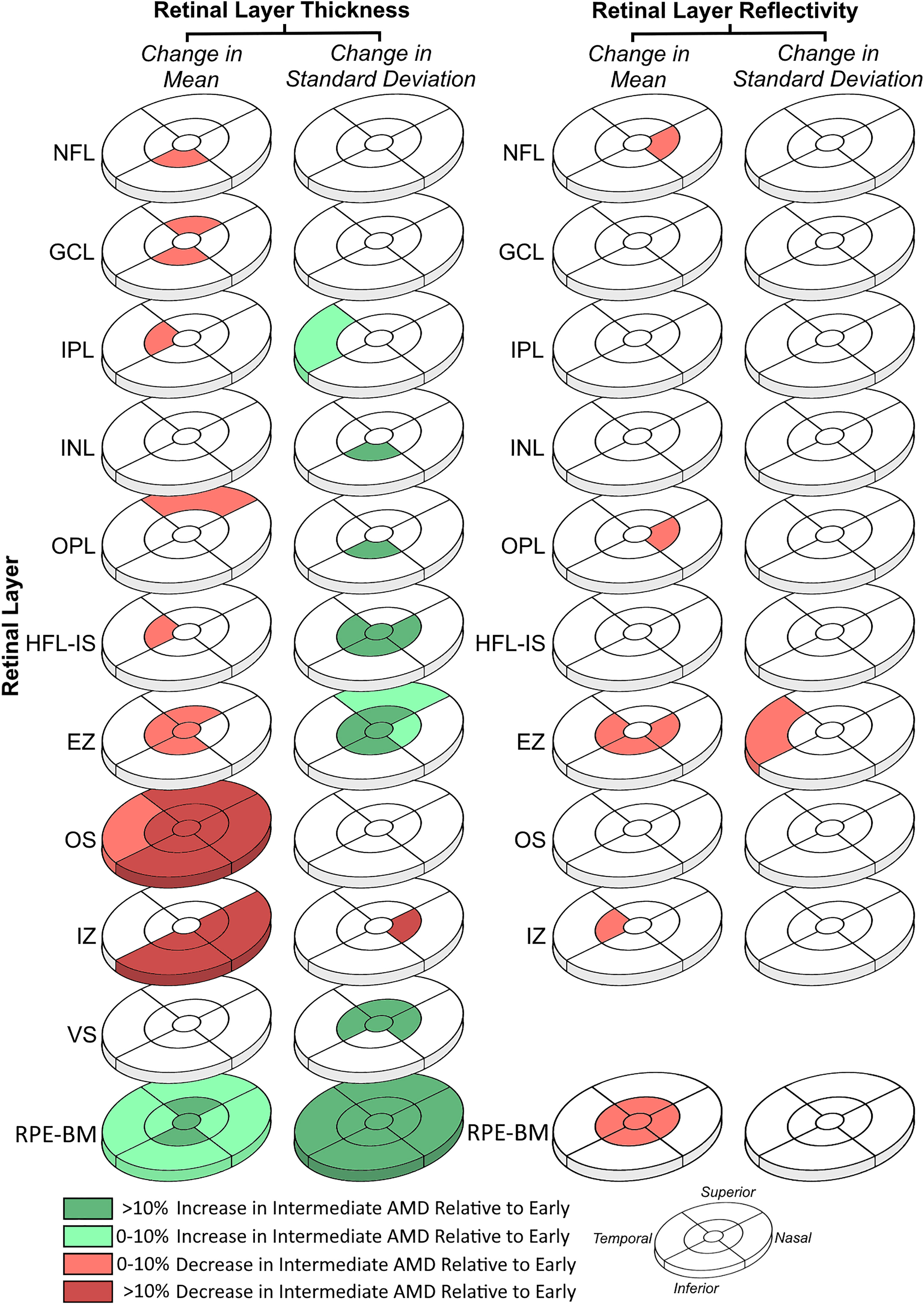
This graphic from the study nicely encapsulates the key findings. Throughout the progression from early to intermediate AMD, researchers observed notable thinning of photoreceptor outer segments thinned notably; within the ellipsoid zone, both the fovea and parafovea showed thinning with a corresponding increase in thickness variability and decrease in parafoveal reflectivity. Photo: Heckenlaible NJ, et al. Ophthalmol Sci. Sept. 4, 2024. Click image to enlarge. |
Automated segmentation of retinal cell layers on OCT makes it possible to observe the morphological characteristics of retinal health during the various stages of age-related macular degeneration (AMD), especially before more evident markers such as drusen and changes in macular pigmentation appear. Limited research has focused on OCT alterations in early AMD; thus, a new study decided to evaluate OCTs from eyes moving from early to intermediate AMD to pinpoint changes in retinal morphology and reflectivity that could act as early indicators of disease progression.
The retrospective cohort study, conducted at Johns Hopkins and UC San Diego, involved 25 patients with AMD over 50 years old. Its authors used a previously validated AI-driven algorithm to automatically segment a total of 50 OCT images (two of the same eye per patient). The scans were analyzed for changes in the mean and standard deviation of cell layer thickness and reflectivity as the disease progressed from the early to intermediate stage.
The results demonstrated that photoreceptor outer segments thinned notably with the progression from early to intermediate AMD. Within the ellipsoid zone, both the fovea and parafovea showed thinning. There was also an observed decrease in parafoveal reflectivity and an increase in the diffuse thickening and thickness variability of the retinal pigment epithelium-Bruch’s membrane (RPE-BM) complex.
All OCT findings corresponded with the known histopathology of the RPE and rod changes in the parafovea in early AMD. “The decreased ellipsoid zone thickness and reflectivity in the parafovea suggests rod photoreceptor degeneration,” the researchers wrote in their paper, published in Ophthalmology Science. “Likewise, the diffuse RPE-BM thickening and variability in the parafovea observed here, while likely primarily due to drusen accumulation, could also reflect the morphologic and molecular heterogeneity that is seen in early AMD.”
The researchers continued, “The mean 3.6 years observed here between early and intermediate stages highlights a period prior to the development of more classic OCT features of AMD.” If disease progression can be recognized sooner through retinal layer changes, this may allow for more tailored and potentially preventative treatment strategies, they suggested, adding that further research involving a larger cohort would help to more decisively establish these imaging findings as reliable markers for risk of early AMD progression.
Heckenlaible NJ, Toomey CB, Handa JT. Optical coherence tomography changes observed during the progression of early age-related macular degeneration. Ophthalmol Sci. September 4, 2024. [Epub ahead of print]. |