 |
|
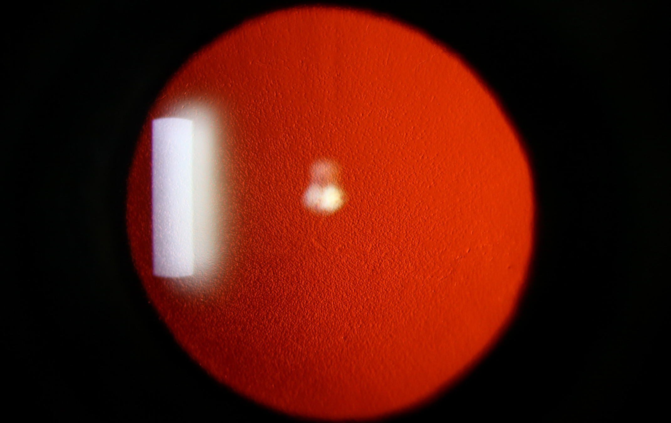
By excluding eyes with subclinical edema, the study confirmed that the smaller, more crowded anterior chamber in FECD was not a result of corneal edema-related posterior expansion of the cornea. Photo: Chris Sindt, OD. Click image to enlarge. |
Fuchs’ endothelial corneal dystrophy (FECD) usually progresses slowly by degrading the endothelial cell layer with eventual stromal and epithelial edema, loss of corneal transparency and diminished vision. A recent study published in Cornea compared several ocular parameters in a large sample of patients with FECD against a group of age- and sex-matched controls. The researchers reported new ocular characteristics in FECD eyes with and without edema. Optical biometry and Scheimpflug imaging established that the anatomic findings in eyes with FECD were not simply due to the larger volume of an edematous cornea but rather unique to eyes with FECD.
A total of 404 eyes (202 eyes with FECD and 202 control eyes) were included in this retrospective chart review. The study included previously described parameters, such as anterior chamber depth, axial length (AL) and spherical equivalent (SE), but it also evaluated additional parameters, including anterior chamber volume, anterior chamber angle, lens thickness, astigmatism, corneal volume and corneal pachymetry performed at different points.
Compared with controls, eyes with FECD had shallower anterior chamber depths, lower anterior chamber volumes and narrower angles. Conversely, the SE before cataract surgery, corneal pachymetry and corneal volume were higher in eyes with FECD. On Scheimpflug imaging analysis, these anatomical differences were present in FECD eyes with and without corneal edema. After adjusting for sex, these differences remained statistically significant. Shorter AL was found to be statistically significant in male eyes but not in female eyes with FECD.
“The association between FECD and eyes with a shorter AL, shallower anterior chamber depth, smaller anterior chamber volume, hypermetropia and narrower anterior chamber angle suggests that FECD occurs more commonly in small eyes. The reason for this is not fully understood,” the study authors wrote in their paper. “A shallow anterior chamber depth and short AL may hasten FECD progression in patients undergoing phacoemulsification or other intraoperative procedures, making the education of patients with FECD regarding their surgical risks an important part of the consent process.”
“The goal of this study was to better define the spectrum of ocular findings in eyes with FECD by using modern optical biometry technology to study a broader range of ocular parameters,” they added. “These findings provide reliable, normative data for future studies that examine surgical, medical and anatomic factors in FECD and for evaluation of the genetic and environmental factors that influence the FECD phenotype.”
Miller DD, Wagner IV, Hasan S, et al. Anatomic characteristics of eyes with Fuchs endothelial corneal dystrophy. Cornea. September 4, 2024. [Epub ahead of print]. |