Age-related reductions in the functioning of the autonomic and cardiovascular systems have been linked with an increased prevalence of cardiovascular diseases and neurological impairments in older people. Several risk factors associated with age-related macular degeneration (AMD) are also implicated in cardiovascular disease, suggesting a shared pathophysiology between the two conditions. Impaired orthostatic hemodynamics can contribute to end-organ damage. The eye, like the brain, is highly vascularized to support the neural retina and thus is potentially sensitive to orthostatic hypotension-triggered blood flow impairment.
 |
|
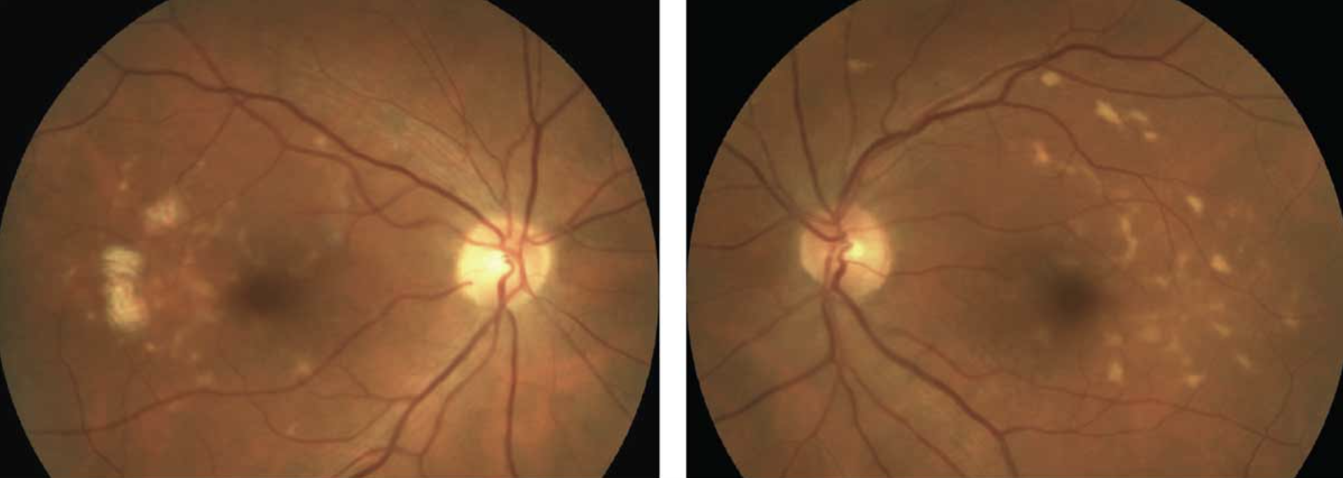
In conjunction with increasing heart rate, the researchers found that cardiac output increased within the first 10 seconds of standing. For participants without AMD progression, this effect was transient and rapidly decreased during the recovery phase. However, in those with progression, the study observed a slower increase in cardiac output in response to standing. Photo: Mohammad Rafieetary, OD, and Roya Attar, OD. Click image to enlarge. |
Researchers at Trinity College in Dublin compared hemodynamic responses to the active stand test in participants with and without AMD progression over a four-year period to determine if indicators of impaired cardiovascular autonomic function were associated with AMD progression during this time. They found significant differences in indicators of impaired hemodynamic responses, particularly in the first 20 seconds post-standing, which could be associated with the pathophysiology of AMD progression. Early orthostatic hemodynamic impairments could offer clues to the pathophysiology of AMD progression and potentially introduce new therapeutic approaches to delay the progression of AMD disease.
The study used a subset of participants from the Irish Longitudinal Study on Ageing, a well-defined sample of community-dwelling adults living with AMD in the Republic of Ireland. Participants with AMD who underwent an active stand test and retinal photographs were included (n = 159; 121 with no AMD progression and 38 with progression). Beat-to-beat hemodynamic data were noninvasively collected during the active stand test, recording systolic blood pressure (BP), diastolic BP, mean arterial pressure (MAP) and heart rate. Cardiac output, stroke volume and total peripheral resistance were derived from these measures. Baseline characteristics were compared between groups with and without AMD progression.
At baseline, increasing age and lower diastolic BP were significantly associated with AMD progression. Mixed-effects models for the period between standing and 10 seconds post-stand revealed significant associations with AMD progression with a steeper drop in diastolic BP and a slower drop in total peripheral resistance. Between 10 and 20 seconds post-stand, AMD progression was significantly associated with a less pronounced reduction in heart rate.
“Together, these findings point toward a possible situation of lower baseline diastolic BP and poor compensatory responses during the early phases of post-orthostasis in participants with AMD progression,” the researchers wrote in their paper for Investigative Ophthalmology & Visual Science. “This could hypothetically reflect a higher risk of transient hypoxia in retinal tissues as a possible underlying pathophysiological mechanism.”
Participants with AMD progression had average diastolic BP levels approximately 5mm Hg lower than participants without progression, suggesting that perfusion of sensitive end-tissues (e.g., myocardium) may be lower and contributing to a higher overall sympathetic tone observed in early hemodynamic responses in the AMD progression group, which could be interpreted as an attempt to compensate for an underlying perfusion problem.
Connolly E, Knight SP, Duggan E, et al. Cardiovascular autonomic function and progression of age-related macular degeneration in The Irish Longitudinal Study of Ageing (TILDA). Invest Ophthalmol Vis Sci. 2024;65(6):24. |