 |
|
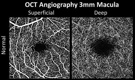
Neither RLRL nor 0.01% atropine appeared to change retinal thickness in premyopic children in this study, but differing effects on the vasculature were noted. Additionally, both treatments prevented myopic shift and axial elongation. Photo: Carolyn Majcher, OD. Click image to enlarge. |
The increasing prevalence of myopia and its associated complications necessitate effective strategies to curb progression. Low-dose atropine and repeated low-level red light (RLRL) have emerged as potential methods for controlling myopia progression in children, though the mechanisms underlying their efficacy remain unclear. A recent study aimed to compare the changes in retinal vascular density (RVD) and retinal thickness (RT) after six months of treatment with RLRL and 0.01% atropine in premyopic schoolchildren. It found that both modalities had similar effects on superficial RVD, while only RLRL led to an increase in deep RVD.
The prospective randomized trial involved 69 premyopic schoolchildren with cycloplegic refraction >-0.75D and ≤0.50D. The children were randomly assigned to either the RLRL or 0.01% atropine group, which had no significant differences in baseline parameters. RVD and RT were measured using swept-source OCT angiography at baseline and after six months. The macular zone was divided into three concentric rings (fovea, parafovea and perifovea) for analysis. Statistical analyses were performed to evaluate the changes in RVD, RT, axial length and cycloplegic refraction between the two treatment groups.
After six months, both RLRL and 0.01% atropine groups showed significant increases in whole, parafoveal and perifoveal superficial RVD. Foveal superficial vessel density remained stable in both groups. In the red light group, whole and perifoveal deep RVD increased significantly while foveal and parafoveal deep RVD remained unchanged. Deep vessel density remained stable in the 0.01% atropine group. Multivariate regression analyses showed that the whole and perifoveal deep RVD increased significantly more in the RLRL group compared to the 0.01% atropine group. When comparing RT between the two groups, the researchers found no significant differences.
In their paper on the study for Translational Vision Science & Technology, the researchers reported that patients on either intervention had significantly increased retinal blood flow compared to placebo. “Subsequently,” they wrote, [this] improved retinal hypoxia in the RLRL group, which may provide a new idea for studying the mechanism of myopia control.”
The researchers also analyzed axial length and refraction data in children treated with RLRL or 0.01%, finding that both interventions effectively prevented myopic shift and axial elongation compared to placebo.
In conclusion, the study authors summarized in their paper, “The present study found that superficial RVD increased with a similar magnitude in the RLRL and 0.01% atropine groups, whereas deep RVD increased only in the former group; there were no significant changes in RT and no documented functional or structural damage in either group after six months of treatment in premyopic children.” They added that further research is warranted to elucidate the long-term effects and optimal treatment regimens for myopia control.
Shang L, Gao S, Wang W, et al. Comparison of changes in retinal vascular density and thickness after using low-level red light and 0.01% atropine in premyopic children. Transl Vis Sci Technol. 2024;13(6):23 |