 |
|
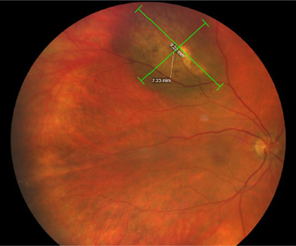
Beware the differences in OCT findings between small, medium and large tumor sizes in choroidal melanoma patients or suspects. Photo: Amy Bade, OD. Click image to enlarge. |
Examining OCT scans for the presence of subretinal fluid can help guide diagnosis and predict prognosis for a wide variety of ocular conditions. A choroidal nevus is a proven risk factor for choroidal melanoma, a condition in which early detection plays a major role in clinical and visual outcomes. Researchers recently determined that choroidal melanoma–associated subretinal fluid has unique features on OCT, including ellipsoid zone loss/disruption, bacillary layer detachment and subretinal hyperreflective material.
The team performed a single-center retrospective review of spectral-domain OCT in treatment-naïve choroidal melanoma with associated subretinal fluid using data from a 12-year period. They reviewed scans from a total of 236 patients (mean age: 65). Not only did the researchers identify several distinctive OCT features among patients with choroidal melanoma, but they also observed a greater extent of subretinal fluid with increasing tumor size.
The most prominent OCT finding among the cohort was ellipsoid zone loss/disruption, observed in 74% of patients. It was more commonly seen in patients with increased subretinal fluid.
Bacillary layer detachment presented in either a classic or floating pattern, the latter of which was more common, observed in 22% of the total cohort and 78% of the patients with bacillary layer detachment. The researchers noted that this could be because intraretinal exudation isn’t often associated with choroidal melanoma. A small percentage of patients (6%) had both classic and floating configurations. Bacillary layer detachment was most common in medium-sized tumors rather than those of small or large size, although the pattern type didn’t seem to be influenced by the size of the tumor. Of all the patients with this finding, 82% had subretinal fluid involving at least one quadrant.
“Tumors with (vs. without) bacillary layer detachment had greater mean presenting tumor thickness (4.4mm vs. 3.6mm) and diameter (13.2mm vs. 11.5mm),” the researchers wrote in their paper published in Retina. Additionally, they identified subretinal hyperreflective material in 36% of patients, which was either heterogeneous, homogenous or both, the first being most common in eyes with bacillary layer detachment vs. those without (58% vs. 20%).
In summary, the researchers concluded, “OCT features of choroidal melanoma–associated subretinal fluid vary by tumor size, with greater subretinal fluid extent in larger tumors, less ellipsoid zone disruption in small tumors and more bacillary layer detachment in medium tumors.
Melis KG, Ferenchak K, Olsen TW, Dalvin LA. Optical coherence tomography findings in choroidal melanoma-associated subretinal fluid. Retina. August 12, 2022. [Epub ahead of print]. |