The glaucomatous lamina cribrosa is subjected to compression/shear forces that distort the axonal fibers within the pores of the structure. These distortions are thought to cause blockage of axonal transport along the intralaminar pathway of the retinal ganglion cell fibers. A research team based in France recently developed semi-automated segmentation software for lamina cribrosa pore detection in vivo, allowing for a 3D reconstruction of the pore pathway from data acquired on SD-OCT to analyze in vivo the path of the pores and better understand the mechanical stress. Changes in pore parameters appeared to be associated with the severity of the glaucomatous optic neuropathy. The lamina cribrosa pores of high-tension glaucoma subjects appeared to be more tortuous than the pores of nonglaucomatous subjects, and the pores of normal-tension glaucoma (NTG) patients were shorter than those of nonglaucomatous subjects.
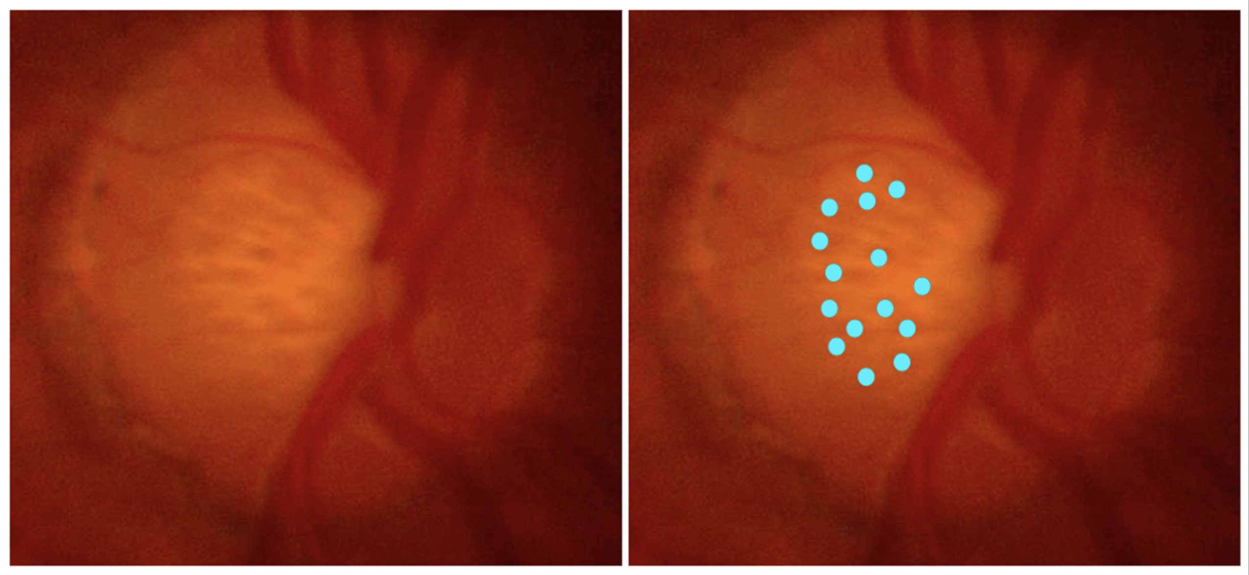 |
| Changes in lamina cribrosa pore parameters appear to be associated with the severity of glaucomatous optic neuropathy. Photo: Jordan JA, et al. Vision. 2024;8(2):24. Click image to enlarge. |
This study, which was published in Journal of Glaucoma, analyzed SD-OCT images of 52 eyes (18 nonglaucomatous, 18 high-tension glaucoma, 16 NTG) of 29 patients over 18 years old with no history of retinal disease or nonglaucomatous optic neuropathy and with an open iridocorneal angle. The team selected only patients with a sufficiently large optic cup to properly visualize the lamina cribrosa. Pores were traced using segmentation software. Pore length, tortuosity and verticality were the three quantitative parameters compared between the three groups.
Pore tortuosity in high-tension glaucoma (1.419) was significantly higher than in nonglaucomatous (1.347) but did not differ from that of NTG eyes. In addition, NTG had significantly shorter pores than the nonglaucomatous group. No difference in pore tortuosity or verticality was found between nonglaucomatous and NTG. Pore verticality and length in high-tension glaucoma eyes did not significantly differ from those of nonglaucomatous eyes and NTG eyes. All of the quantitative parameters measured were not correlated with age but were associated with glaucoma severity (visual field index, mean deviation, retinal nerve fiber layer; RNFL, ganglion cell complex thickness; GCC), except for pore verticality, which was not correlated with RNFL.
“As it appears correlated to GCC damage, the increase in pore tortuosity could be altered as early as, or even precede, the first axonal lesions and thus constitute a predisposing factor for glaucoma,” the study authors wrote in their paper. “Further studies are needed to understand the chronological sequence of the changes in these new parameters and the first axonal lesions related to glaucoma and to clarify the role that this new imaging technique could play in the diagnosis and monitoring of glaucoma patients.”
Still, the study noted that the work was based on the analysis of a small sample of the population of interest. As a result, the statistical analyses lacked power, and further work on larger numbers is still needed. Because of the overall hyperreflectivity of the images and the presence of intralaminar defects probably related to fibrous remodeling of the lamina cribrosa, fewer pores could be traced correctly in the two groups of glaucoma patients. The team then decided to limit the evaluation to between five and 10 pores for each eye. Another well-known and unavoidable limitation of tomographic imaging of the lamina cribrosa was that the images obtained on OCT allow only partial visualization of the laminar tissue. Notably, the size of the optic cup has been identified as a risk factor for glaucoma. So, these nonglaucomatous patients analyzed, although totally normal regarding glaucoma diagnostic criteria, were more susceptible to future glaucoma than the general population.
Bastelica P, Labbé A, Hamard P, et al. Three-dimensional microarchitecture of lamina cribrosa pores in high and normal tension glaucoma using optical coherence tomography. J Glaucoma. September 11, 2024. [Epub ahead of print]. |