 |
|
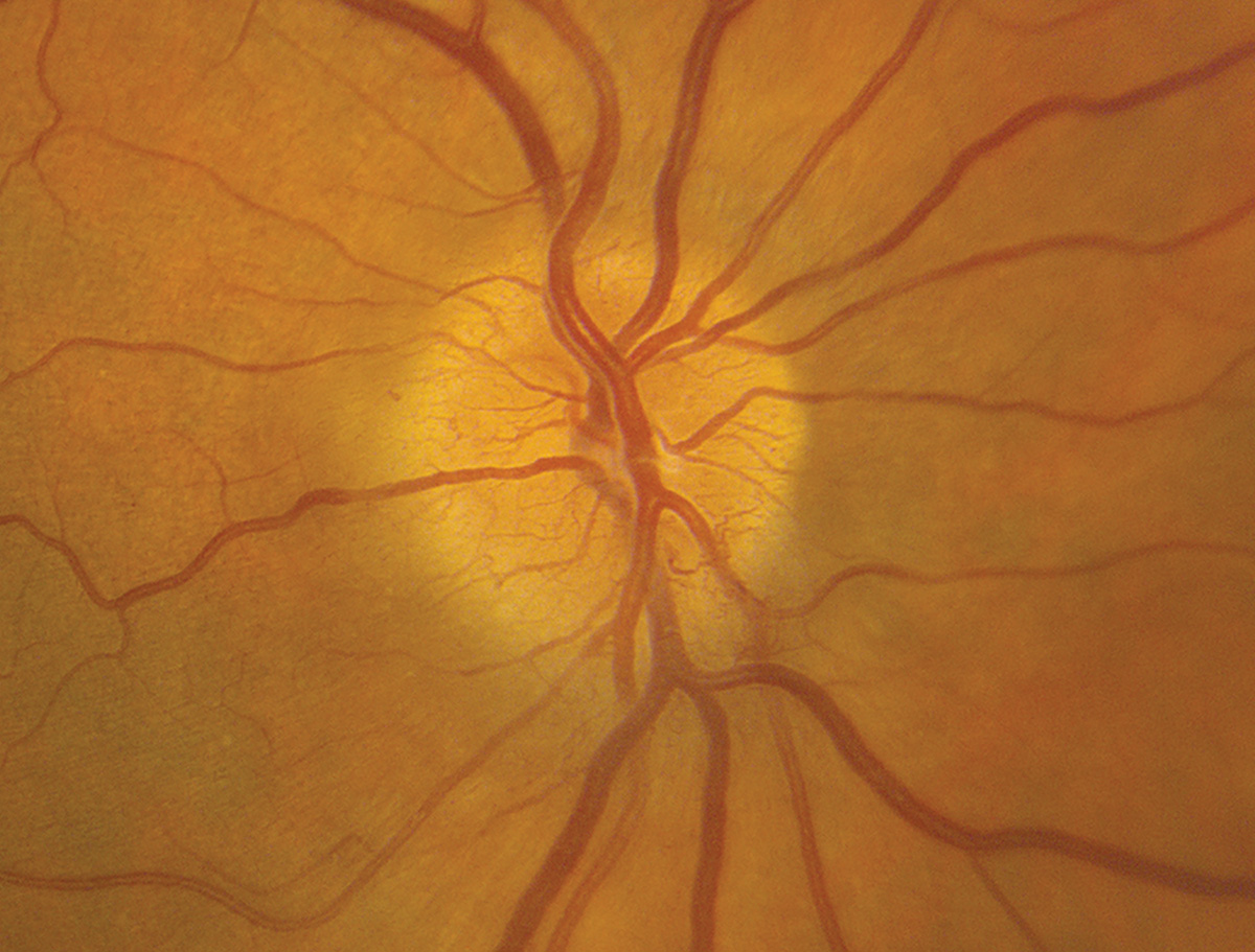
Hyperemia was the most common fundus feature identified in acute NAION patients in this study. While no single fundus feature correlated to VF loss, combining multiple features resulted in an accuracy of 73.6% in classifying the level of VF loss. Photo: Alison Bozung, OD. Click image to enlarge. |
While numerous studies have explored the correlation between fundus features and visual loss in macular disorders and glaucoma, there is a lack of comparable research focused on non-arteritic anterior ischemic optic neuropathy (NAION). A recent study, whose results were presented last week at ARVO in Seattle, aimed to fill this gap by investigating fundus features of the optic nerve head and peripapillary retina and their correlation to visual field (VF) loss severity in acute NAION.
The prospective study analyzed 728 fundus photos of eyes with acute NAION from the Quark trial, an investigational drug study for NAION that launched back in 2016. Participants were assessed within 14 days from the onset of vision loss. Fundus features related to optic nerve head swelling were identified and segmented, and images were graded based on the level of vessel obscuration for each optic nerve head quadrant. VF loss severity was categorized as moderate (>-12dB), severe (-12dB to -20dB) or profound (<-20dB) based on average total deviation.
Among the 728 fundus images, 86.2% had optic nerve head hyperemia, 77.9% had hemorrhages, 27.4% had cotton wool spots, 21.1% had pallor and 14.5% had peripapillary wrinkling. The VF data (available for 679 eyes) showed a mean total deviation of -18.5 dB (+/-7.5). Of these, 21.1% had moderate VF loss, 39.6% had severe VF loss and 39.3% had profound VF loss.
Although individual features revealed only weak associations with VF loss severity (total swelling: 0.13; total elevation: 0.13; wrinkles: 0.11; pallor: 0.09; hyperemia: -0.11), considering them in combination "resulted in an accuracy of 73.6%” at classifying the level of VF loss, the researchers wrote in their ARVO abstract on the study.
These findings demonstrate the importance of assessing fundus features, particularly optic nerve head swelling, to help predict and understand the progression and severity of acute NAION. Further investigation will be needed to explore the evolution of structural features over time and their relation to visual field outcomes.
Original abstract content ©2024 Association for Research in Vision and Ophthalmology.
Woods B, Szanto D, Stern L, et al. Fundus features relate to severity of visual field loss in acute non-arteritic anterior ischemic optic neuropathy (NAION). ARVO 2024 annual meeting. |