 |
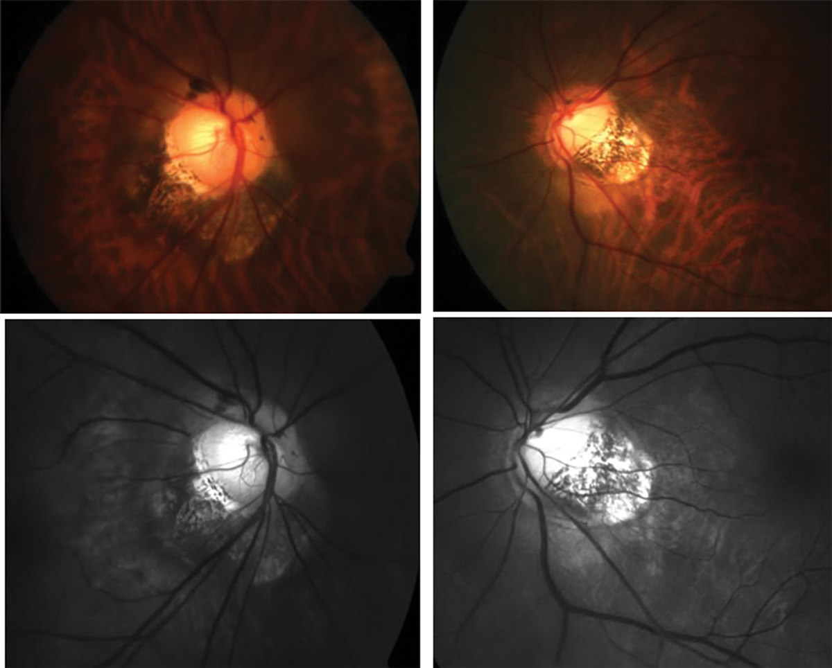
| While the development of myopic macular retinoschisis is multifactorial, this study found that patients with higher depth of the wide and narrow primary macular staphyloma were at greater risk. Photo: Andrew Rouse, OD. Click image to enlarge. |
A recent cross-sectional study investigated how varying dimensions of primary macular staphylomas in highly myopic patients influence the development of myopic macular retinoschisis. The goal of the research was to identify the structural variables that may predict the likelihood of this vision-threatening complication, which affects between 8% and 34% of high myopes.
The study reviewed data of 52 patients (69 eyes) with high myopia and wide or narrow macular staphyloma from a single university hospital in Spain (mean patient age: 54 years). Swept-source OCT and ocular ultrasonography were employed to measure the depths of macular staphylomas and assess the presence of myopic macular retinoschisis.
Myopic macular retinoschisis was detected among nine of the 69 eyes, with the remaining 60 eyes showing no macular pathology. Most of the cohort (61 eyes; 88.4%) presented with a wide primary macular staphyloma, while a smaller percentage (9 eyes; 11.59%) had a narrow macular staphyloma.
The findings highlighted a notable correlation between the depth of staphylomas and the occurrence of myopic macular retinoschisis. Specifically, deeper and more pronounced staphylomas were associated with a higher incidence of retinoschisis.
“Compared to eyes without disease, eyes with myopic macular retinoschisis showed a significant associated with vitreomacular traction, retinal arteriolar traction, greater axial length and higher depth of the wide and narrow primary macular staphyloma,” the researchers reported in their paper, published last week in the Retina journal. Additionally, they noted that “all patients with myopic macular retinoschisis presented a multiple columnar structure between the inner and outer retina with the macula at the deepest part of the staphyloma.” The multivariate analysis corroborated these findings.
This study has several limitations, including a small sample size and cross-sectional design. Also, measurements of posterior staphyloma depth were conducted by averaging four assessments performed by an ophthalmologist, raising potential concerns about measurement consistency that could be explored in future research to evaluate reproducibility. Lastly, while myopic macular retinoschisis can manifest internally or externally (based on which area of the neurosensory retina the schisis impacts), this study could not classify the types due to limited cases.
The researchers concluded that despite these limitations, “this study provides evidence that myopic macular retinoschisis seems to be multifactorial.” They added that to their knowledge, “this is the first study to demonstrate that highly myopic eyes with a greater depth of wide and narrow primary macular staphyloma are at higher risk of developing myopic macular retinoschisis.”
| Click here for journal source. |
García-Ben A, Martín MF, Pascual NO, et al. Relationship between the height of wide and narrow primary macular staphyloma and myopic macular retinoschisis. Retina. October 2, 2024. [Epub ahead of print]. |