Although fluorescein angiography has been the preferred imaging method for diabetic retinopathy, its invasive nature has led researchers in the field to explore alternative options for capturing capillary nonperfusion in the superficial capillary plexus and deep capillary plexus. Increasingly, OCT-A vascular metrics are being used to assess diabetic retinopathy (DR) severity, but there’s no universal standard for the most appropriate metrics to use as biomarkers of progression.
A new study specifically looked at the characterization of non-proliferative diabetic retinopathy (NPDR) progression using OCT-A metrics along with microaneurysm formation and disappearance rates in fundus images. Researchers found that skeletonized vessel density (SVD) and perfusion density (PD) do in fact detect capillary NPDR. Initially, the capillary nonperfusion involves the superficial capillary plexus (SCP) and later progresses to the deep capillary plexus (DCP).
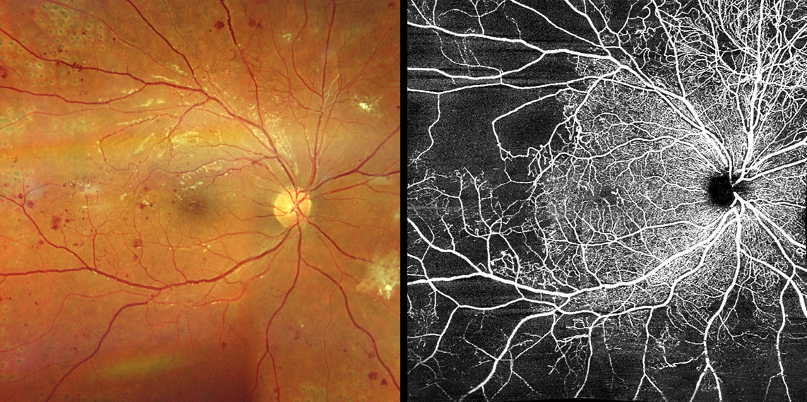 |
| Using certain OCT-A metrics, researchers detected significant capillary nonperfusion in the inner ring area of the superficial capillary plexus for patients in the earliest stage of NPDR but found capillary closure in the superior and deep capillary plexus for patients in both the mild NPDR and moderate or moderately severe NPDR categories. Photo: Carolyn Majcher, OD. Click image to enlarge. |
A total of 122 eyes were included in this study, which was a post-hoc analysis of a prospective longitudinal cohort study known as CORDIS, with a two-year duration. When categorized by severity as defined by ETDRS staging, eyes graded as ETDRS level 20 showed significant capillary nonperfusion predominantly in the inner ring area in the SCP, whereas eyes graded as ETDRS level 35 and ETDRS levels 43 and 47 showed significant capillary nonperfusion in both the SCP and DCP in both inner and outer rings. This confirms previous reporting by the group that demonstrated progressive decentralization of the ischemia and involvement of more peripheral regions of the retina. Rates of progression changes were found predominantly in the DCP for SVD and PD and were better identified in the outer ring area.
Microaneurysm turnover also contributed to the characterization of NPDR progression by discriminating ETDRS level 35 from ETDRS levels 43 and 47, which could not be achieved using only OCTA metrics. The authors say microaneurysms have been shown to be preferentially located in abnormally dilated shunt vessels and have been proposed to be particularly relevant to NPDR progression.
Despite the limitations of the study, including its restrictive inclusion criteria that was focused on a metabolically well-controlled population with a higher percentage of men, the authors state in their paper on the work that the combination of OCT-A and microaneurysm turnover is clinically relevant. “This study offers data that support a coherent interpretation of the progression of the microvascular disease component of NPDR,” they wrote. “It indicates that OCT-A metrics performed in both central 3x3 mm and 6x6 mm regions of the retina are not sufficiently discriminative of ETDRS levels, and the additional calculation of microaneurysm turnover offers necessary and complementary information that appears to accurately identify the different severity grades of NPDR in the clinical setting.”
Marques IP, Ribeiro ML, Santos T, Reste-Ferreira D, Mendes L, Martinho AC, Santos AR, Figueira J, Lobo C, Cunha-Vaz J. Patterns of progression of nonproliferative diabetic retinopathy using non-invasive imaging. Transl Vis Sci Technol. 2024;1:13:5:22. |