 |
|
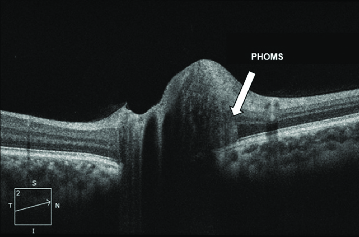
New research suggests that idiopathic intracranial hypertension is more pronounced in patients previously diagnosed with isolated PHOMS, necessitating ongoing assessments. Photo: Bassi ST, Indian J Ophthalmol 2023;71:3552-7. Click image to enlarge. |
A new study sought to delineate the clinical characteristics and imaging features of peripapillary hyperreflective ovoid mass-like structures (PHOMS) to assist in reliably distinguishing them from other optic nerve head anomalies, such as optic disc drusen.
The retrospective, observational study was conducted at Rothschild Foundation Hospital, including patients seen within a four-year window with an elevated optic disc with blurry margins (EODBM) and PHOMS. Cases with papilledema with Frisén grade ≥3, optic neuritis, ischemic optic neuropathy and compressive optic neuropathy were excluded. Data collection involved OCT imaging and analysis of the PHOMS' volumes across different eye quadrants, coupled with monitoring for signs of idiopathic intracranial hypertension (IIH) and papilledema.
The study cohort comprised 308 eyes of 154 patients (78% female; mean age: 29 years). PHOMS were primarily associated with cases of IIH (38.3%), with other etiologies including isolated IIH (35.7%), posterior uveitis (11%), optic disc drusen, (10%) and tilted optic disc (5%). The finding was most prevalent in the nasal region. In their paper on the study, the researchers pointed out, “The location of PHOMS in the superior and/ or inferior quadrant was significantly associated to IIH or optic disc drusen, while their presence in the temporal or nasal sector was strongly associated to isolated lesions.” Based on this, the authors suggest that a vertical distribution of PHOMS (superior and inferior regions) should raise suspicion for papilledema or another pathological configuration of the optic nerve head.
In the 83% of the cohort with MRI data, the researchers found that more than half of scans were interpreted as consistent with IIH, although only 39.7% of these patients had confirmed IIH with poor sensitivity (44.5%) and sensibility (55.5%). A recent study they cited in their paper found that radiologic signs of IIH had 74.8% sensitivity and 94.7% specificity, supporting the idea that “IIH diagnosis should always rely on a strong clinical suspicion and a confirmed elevated pressure measurement, rather than on the MRI findings alone.”
The researchers elaborated in their paper that PHOMS are “often responsible for misleading ophthalmologists to believe that they are the sign of an ongoing papilledema.” When evaluating an asymptomatic patient for EODBM, they urge physicians to remember “that PHOMS can be an associated sign of IIH, but can also represent isolated variation of normal anatomy, even in the absence of a tilted disc.”
Lastly, the study authors noted that upon revisiting their initial diagnosis of isolated PHOMS in three patients over 12 months of follow-up, they noticed that IIH became more evident and both peripapillary RNFL and PHOMS volume increased throughout this time. This highlights that while isolated PHOMS appears to be benign and pose no risk to vision, it is still crucial to monitor patients consistently.
In conclusion, the authors wrote, “Isolated PHOMS should be considered as a distinct entity. In asymptomatic patients, PHOMS should be carefully studied. Nasal or temporal location, small volume and stable aspect over the course of weeks or months are so suggestive of this entity.” They speculate, “This strategy would considerably reduce the impact on patients’ anxiety and morbidity.”
| Click here for journal source. |
Maalej R, Bouassida M, Picard H, Vignal Clermont C, Hage R. Are PHOMS (peripapillary hyperreflective ovoid mass-like structures) with an elevated optic disc still a diagnosis dilemma? Ophthalmology. September 13, 2024. [Epub ahead of print]. |