Current technology for measuring axial length included optical biometry and ocular ultrasound. An intriguing new study explores how OCT’s high-resolution cross-sectional images hold potential in predicting axial length (AL) and providing further structural information that has yet to be explored. Korean researchers in Seoul used a deep learning model that analyzed horizontal and vertical OCT images as dual inputs to detect the dome-shaped macula and predict AL accurately.
A total of 9,064 eyes of 5,349 patients (total image number of 18,128) were included. The average AL of the eyes was 24.35mm. AL measurement was taken using IOLMaster 700 (Carl Zeiss Meditec), and an OCT image was taken by Spectralis OCT (Heidelberg Engineering) within three months prior to AL measurement. A horizontal and vertical linear OCT scan of 8.8mm centered onto the fovea acquired in high resolution modality with a 100 auto real time resolution was carried out using Spectralis HRA + OCT. The OCT images were classified based on the presence of macular abnormalities, which included epiretinal membrane (ERM), age-related macular degeneration (AMD), central macular edema (CME), macular hole or other macular abnormalities.
Deep learning models predicted AL with mean absolute error (the mean of the difference between model prediction value and the actual value) and R-squared value (another measure of accuracy) of 0.592mm and 0.847mm, respectively, in the internal test set (1,824 eyes of 1,070 patients). In the external test set (171 eyes of 123 patients), the deep learning models predicted AL with MAE and R2 of 0.556mm and 0.663mm, respectively. Regarding error margins of ±1.0mm, ±2.0mm and ±3.0mm, the dual-input models showed accuracies of 83.5%, 98.1% and 99.5%, respectively, in the internal test set, and 85.4%, 99.4% and 100%, respectively, in the external test set.
 |
|
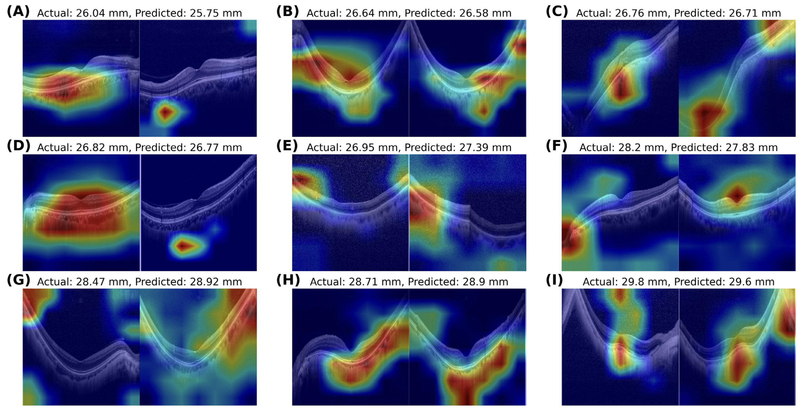
This novel model demonstrated impressive performance and could possibly be used not only for accurate AL prediction based on OCT images but also for investigating how retinal and choroidal structures are related to AL elongation. This image from the study—a heatmap analysis of myopic eyes with axial length greater than 26.0mm—shows the actual and OCT-predicted values for nine subjects in the study. Photo: Oh R, et al. Transl Vis Sci Technol. 2024;13(9):14. Click image to enlarge. |
“Our study samples encompass not only normal retinal images but also a diverse range of pathological conditions,” the study authors noted in their paper. “This inclusion of various diseases enhances the real-world relevance and applicability of our findings, as it accounts for the complexity and heterogeneity of retinal fundus lesions that may impact eyeball elongation.”
The analysis was based solely on horizontal and vertical cross-sectional images, without incorporating macular volume scan images. The team suggested that future studies should consider incorporating macular volume scan images to improve the accuracy of prediction.
Perhaps one day, OCT devices may eventually allow doctors to measure axial length directly.
| Click here for journal source. |
Oh R, Kang M, Ahn J, et al. Prediction of axial length from macular optical coherence tomography using deep learning model. Transl Vis Sci Technol. 2024;13(9):14. |