The precise prevalence of the compound form of wide macular posterior staphyloma and its clinical features compared to its primary form have not been reported yet, although the compound form seems to account for a considerable proportion of cases. Researchers in Seoul, South Korea, recently compared the ocular features of highly myopic eyes with posterior staphyloma of wide macular type according to their morphological complexity. They were classified into the primary (Curtin type I) and compound (Curtin types VI to X) forms based on the configuration within the staphyloma. The compound form of wide macular posterior staphyloma had more severe myopic macular changes and a worse visual prognosis compared to the primary form, and these were associated with more structural deformation in the posterior eyeball.
 |
|
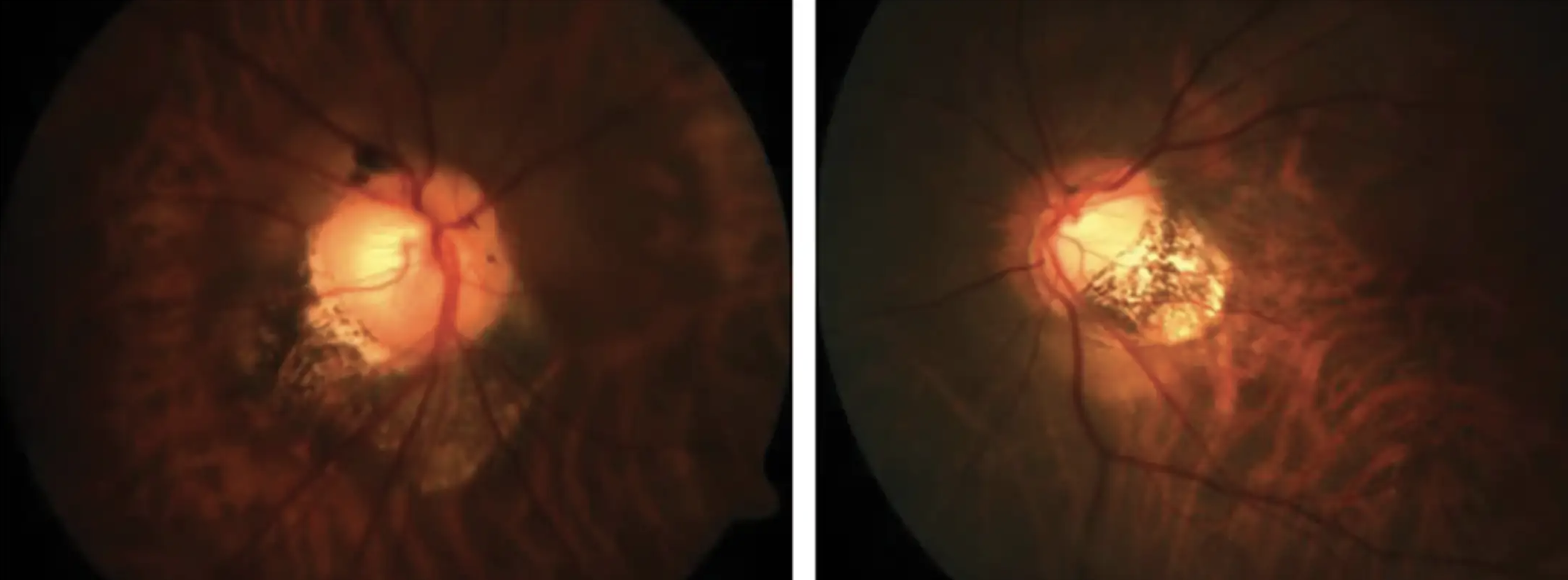
Eyes with compound wide macular posterior staphyloma had higher grades of myopic macular degeneration and higher frequency of neovascularization and foveal change associated with myopic macular traction. Photo: Andrew Rouse, OD. Click image to enlarge. |
A total of 154 eyes (103 patients) with primary type and 65 eyes (49 patients) with compound type were included. The grades of myopic maculopathy and the thicknesses of the choroid and sclera were compared between each group. Eyes with compound wide macular posterior staphyloma had worse visual acuity and greater axial length than those with the primary form. The compound type also had a higher grade of myopic macular degeneration and a higher frequency of lamellar or full-thickness macular holes associated with myopic traction (21.5% vs. 10.4%) and active or scarred myopic choroidal neovascularization (33.8% vs. 20.1%).
On swept-source OCT, eyes with compound wide macular posterior staphyloma had significantly thinner choroid and sclera. The study authors noted, “Although this new classification is more objective and simpler to categorize the morphology of posterior staphyloma, the detailed frequency of the compound-form wide macular posterior staphyloma based on new imaging modalities such as ultra-widefield fundus imaging and widefield OCT has not been reported.”
“Worse visual prognosis associated with more severe structural deformation in eyes with the compound form of wide macular posterior staphyloma implies that it may be a more advanced form, and more attention should be paid to the development and progression of myopic macular complications,” they wrote in their paper. “Choroidal and scleral thicknesses were more decreased in the compound form, which may imply that the biomechanical properties of the sclera are more altered than those of the primary form.”
Yoon CK, Lee EK, Bae K, Park UC. Clinical features of primary and compound forms of wide macular posterior staphyloma in high myopia. BMC Ophthalmol. 2024;24:246. |