 |
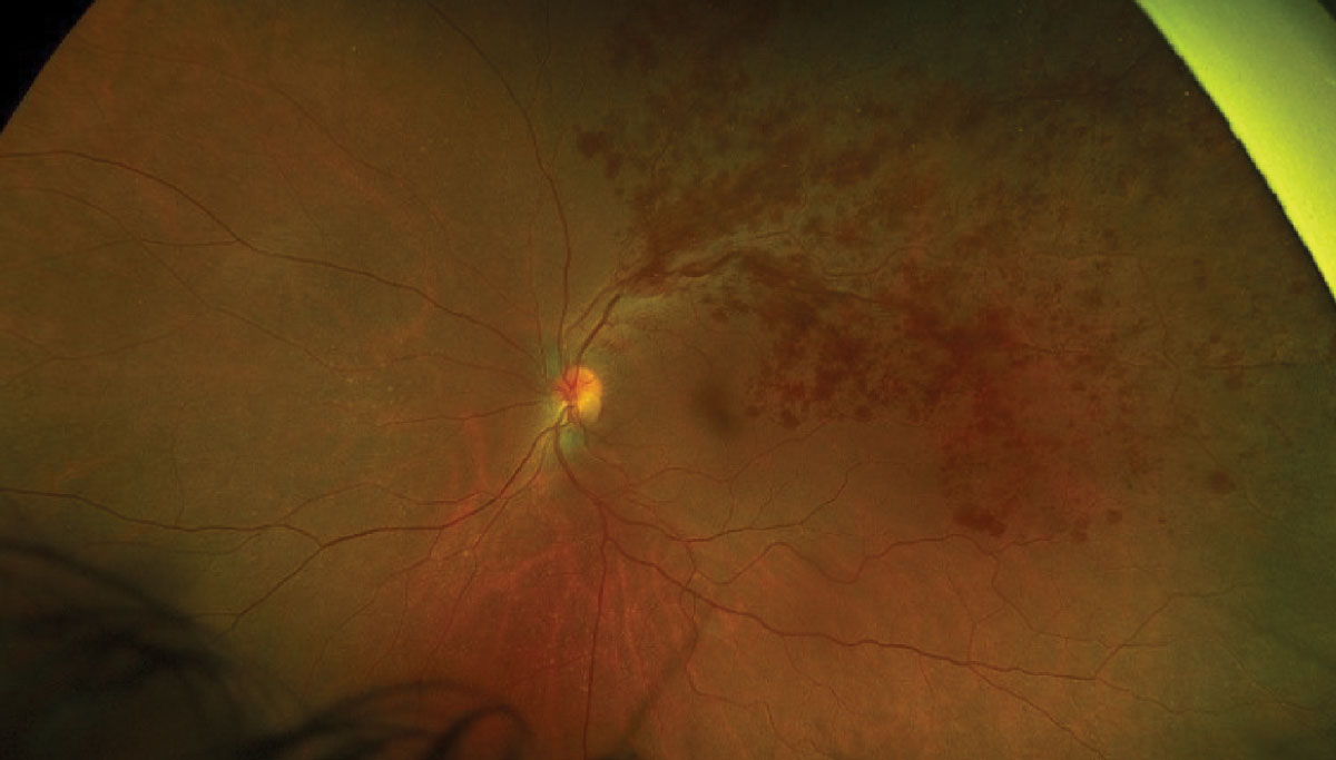
| Recent findings implied that both green and red channel hemorrhage measurements can provide some indication of nonperfusion areas, with greater value attributed to peripheral hemorrhages. Photo: Julie Torbit, OD, and Brad Sutton, OD. Click image to enlarge. |
The responsibility of establishing retinal perfusion status holds great importance in patients with branch retinal vein occlusion (BRVO), as the presence and extent of ischemia are associated with a worse prognosis. Currently, assessment of nonperfused areas in retinal vascular diseases such as BRVO primarily relies on fundus fluorescein angiography (FFA), which is invasive, time consuming and only offers limited visualization of the peripheral retina.
Ultra-widefield (UWF) fundus imaging is more readily available outside of retina subspecialty clinics and has the advantages of noninvasiveness and a nearly 200° field of view, enabling rapid and comprehensive imaging of vascular tortuosity and retinal hemorrhage in the affected area. The Optos imaging system uses red and green laser wavelengths to capture ultra-widefield pseudocolor images of the retina. Green-channel imaging predominantly highlights the retina and its vascular structures, while red-channel imaging provides visualization of deeper structures such as the deeper retina, RPE and choroid.
Researchers in China recently explored the distribution of hemorrhage on the red and green channels of UWF imaging in patients with ischemic and nonischemic BRVO, while examining its association with nonperfusion areas. They found that the detection of hemorrhages on the red and green channels using easily accessible UWF imaging allowed for an initial prediction of nonperfusion areas, particularly in the peripheral region.
This retrospective cross-sectional study, published in Retina, included 96 patients, 46 with ischemic BRVO and 50 with nonischemic BRVO. The researchers further divided the retina into posterior and peripheral areas to assess the association between hemorrhage and nonperfusion areas in different regions. By selecting the panretina, posterior area, and peripheral area, the team used the "measure" option on the Optos 200TX to calculate the percentages of green channel hemorrhage, red channel hemorrhage and nonperfusion area in each region.
Ischemic BRVO showed significantly higher percentages of green channel hemorrhage percentage and red channel hemorrhage than nonischemic BRVO in the peripheral regions, while no significant differences were observed in the panretinal and posterior area. Significant correlations were found between percentage of nonperfusion area in the panretinal and peripheral areas and the corresponding percentages of green and red channel hemorrhages. However, no significant correlation was observed between posterior nonperfusion area percentage and posterior green channel and red channel hemorrhages. Additionally, peripheral green and red channel hemorrhages were related to panretinal percentage of nonperfusion area.
“The observation of nonperfusion areas in the periphery, which is challenging under standard FFA, holds significant importance for predicting neovascular complications,” the study authors wrote in their paper. “This early identification can alert physicians about the occurrence of these nonperfusion areas, potentially influencing patient management strategies.”
Sun G, Wang X, Yi z, et al. Relationship between retinal hemorrhage on green and red channels of ultra-widefield fundus images and retinal perfusion in acute branch retinal vein occlusion. Retina. December 28, 2023. [Epub ahead of print]. |
