 |
|
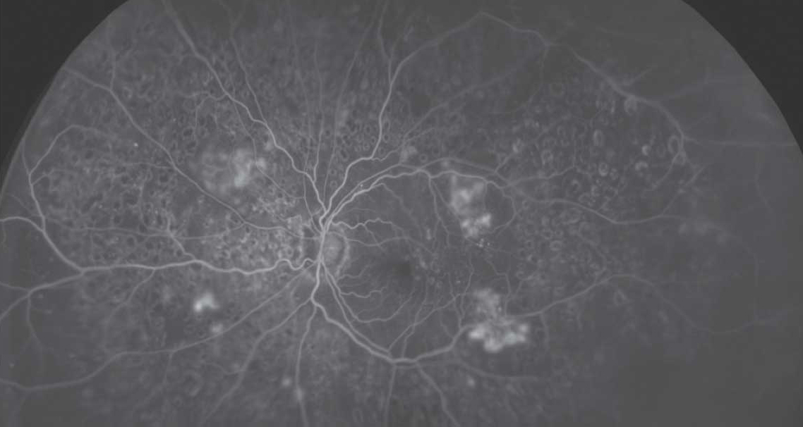
Predominantly peripheral lesions, especially severe hemorrhages/microaneurysms and intraretinal microvascular abnormalities, showed a strong association with the progression of diabetic retinopathy in this study, emphasizing the value of using ultra-widefield FA imaging to assess these patients. Photo: Optos. Click image to enlarge. |
Advancements in retinal imaging technology now enable the evaluation of a significantly larger section of the retina, enhancing optometrists’ ability to assess progression risk in diabetic retinopathy (DR). A 2022 observational longitudinal study called Protocol AA, led by the DRCR Retina Network, aimed to identify eyes at elevated risk of DR progression using ultra-widefield (UWF) imaging to detect the presence or absence of predominantly peripheral lesions. Its findings indicated that while lesions observed in color photographs did not correlate with an increased likelihood of disease worsening, their presence in UWF fluorescein angiography (UWF-FA) did link to a notably higher risk of disease progression.
A group of ophthalmologists from the DRCR network recently performed a post hoc secondary analysis on data from Protocol AA to explore further how lesion location and severity on UWF imaging might more precisely predict progression risks.
“As shown by Protocol AA and independent reports from multiple groups, UWF imaging documents retinal lesions that develop in the retinal area not captured by standard retinal imaging,” they wrote in their paper, published recently in the journal Retina. “This study evaluates specific DR lesion types outside the Early Treatment Diabetic Retinopathy Study (ETDRS) 7-fields that may further refine the risk of progression.”
The post hoc secondary analysis included 544 eyes of patients with nonproliferative DR and an ETDRS-DRSS score between 35 and 53 who were followed for four years. Researchers used both UWF color and UWF-FA images to assess lesion impacts beyond the ETDRS standard fields. Lesions were categorized based on their visibility in UWF images (with and without a template mask overlay) to determine their influence on progression severity.
The findings highlighted that severe lesions outside the traditional ETDRS fields, especially on UWF-FA images, significantly correlated with an increased risk of disease progression. Greater risk of worsening over four years was associated with certain lesion types, including severe hemorrhages/microaneurysms and intraretinal microvascular abnormalities visible on UWF-FA. “This finding suggests that failing to identify eyes that have peripheral DR lesions may lead to underestimation of risk of DR worsening, and that identification of lesions throughout the entire retina is important to stratify risk more accurately for progression over time,” the researchers explained in their Retina paper.
In conclusion, they noted this data demonstrates that “DR lesions identified in UWF-color or UWF-FA images are associated with similar risks for disease worsening, regardless of their placement predominantly within the ETDRS fields, predominantly outside these fields, or uniformly distributed across the retina.” These results align with foundational studies using fluorescein angiography and newer UWF-FA data, the researchers pointed out, which indicate that clinical and vascular lesions in DR predominantly appear in the mid-peripheral retina.
“Although the optimal method of disease risk assessment on UWF imaging still needs to be determined,” the study authors argue, “the detailed grading information from UWF-color, UWF-FA and nonperfusion assessments of the AA data set will allow further analyses to potentially improve our ability to identify diabetic eyes at increased risk of disease worsening.”
| Click here for journal source. |
Silva PS, Liu D, Aiello LP, Melia M, Sun JK, for the DRCR Retina Network. Diabetic retinopathy lesion types and distribution on ultrawide field imaging and the risk of disease worsening over time. Retina. September 12. 2024. [Epub ahead of print]. |