 |
|
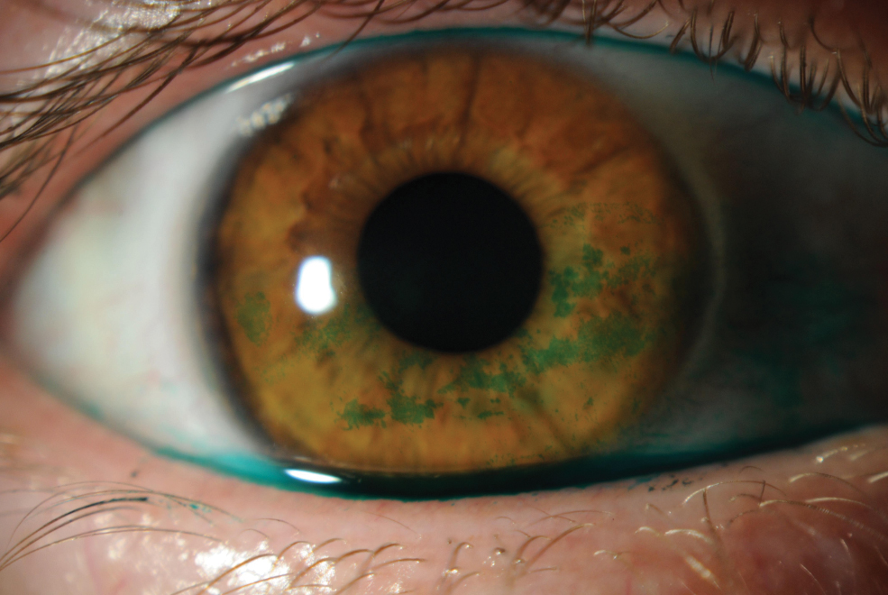
This study explained how applying a lissamine green strip five seconds after wetting it, and then applying the same strip one minute after to the ocular surface was the most optimal way to observe the bulbar conjunctiva, but the amount of dye used for each patient was not measured. Photo: Rebecca Rojas, OD. Click image to enlarge. |
The method of applying and assessing lissamine green staining was thoroughly detailed in the 2017 Tear Film and Ocular Surface Society’s Dry Eye Workshop II (TFOS DEWS II) report. Although the report specified exactly how long to wait after wetting a lissamine green strip with saline—five seconds—it recommended that observation should take place between one and four minutes.1 This broad timeframe could be optimized to maximize the conjunctival staining observed.
Researchers from the United Kingdom attempted to develop a lissamine green staining technique that could provide the best observational results. The team devised five application methods. One included applying the strip immediately after wetting. Another included applying the strip five seconds after wetting. Similarly, the third method included applying the strip 10 seconds after wetting. Additionally, consecutive single applications of sodium fluorescein followed by lissamine green were applied five seconds after wetting, and each were administered one minute apart. The final method included two applications of the same lissamine green strip five seconds after wetting, both one minute apart as well.
After carefully designing each application method, the researchers recruited 22 subjects for their study. Each subject was randomly assigned an application method. After staining, punctate spot count and lissamine green staining intensity were assessed using slit lamp photography at 30, 60, 90 and 400 seconds.2
“Values for punctate spot count and lissamine green staining intensity varied significantly between the different application methods,” reported the researchers in their study. According to their findings, two applications of the same lissamine green strip five seconds after wetting, both one minute apart, provided the best results for both punctate spot count and lissamine green staining intensity. All other methods were significantly lower. Although, applying the strip five or 10 seconds after wetting was more effective than immediate application when observing for punctate spots. However, the five-second method outperformed the 10-second and immediate methods with regard to lissamine green staining intensity.
“It is also recommended that to observe the greatest extent of bulbar conjunctival staining spot count and staining intensity, the bulbar conjunctiva should be viewed immediately after lissamine green application,” mentioned the researchers in their study. Their study provides an optimized method for lissamine green staining that further strengthens what’s instructed in the DEWS II report. But they believe more work needs to be done, especially since there method of combining fluorescein and lissamine green dyes did not enhance the visualization of the ocular surface.
1. Wolffsohn JS, Arita R, Chalmers R, et al. TFOS DEWS II diagnostic methodology report. The Ocular Surface 2017;15:539–74. 2. Ghorbani-Mojarrad N, Vianya-Estopa M, Martin E, et al. Optimizing the methodology for the assessment of bulbar conjunctival lissamine green staining. Optometry and Visual Science. August 26, 2024. [Epub ahead of print]. |