 |
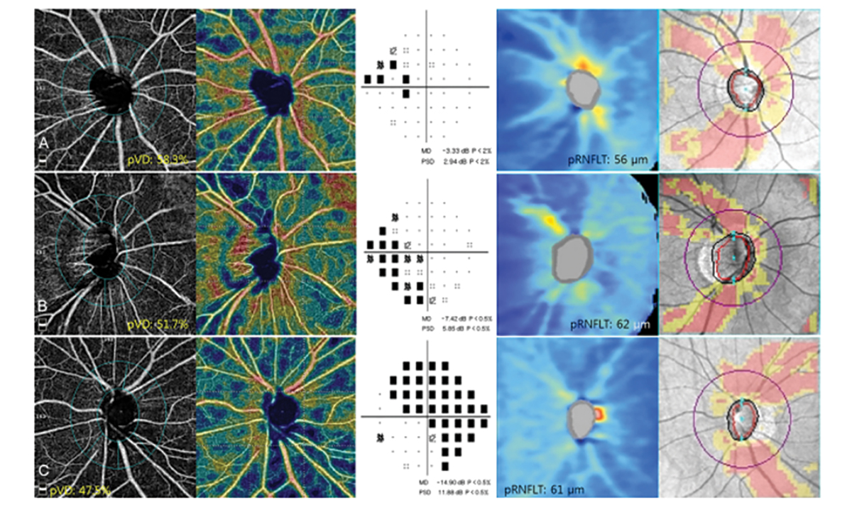
| This study observed that vessel density measurements in peripapillary sectors are lower in eyes with a tilted disc. Photo: Andrew Rouse. Click image to enlarge. |
To properly differentiate glaucomatous from healthy eyes, it is important to determine how optic nerve head deformation alters peripheral vessel density and to understand the effect of this deformation on the reproducibility of OCT angiography (OCT-A) measurements.
Peripheral vessel density measured by this technology has shown good short-term and long-term reproducibility in eyes with and without glaucoma, as well as in eyes with other optic nerve diseases such as optic nerve atrophy and disc edema. However, a recent study determined that disc tilt was independently associated with lower peripheral vessel density. It also found that the long-term reproducibility of OCT-A peripheral vessel density measurements was lower in eyes with a tilted disc.
This retrospective, observational case-control study included 70 healthy eyes. Average and eight-sector peripheral vessel densities and peripapillary retinal nerve fiber layer (RNFL) thicknesses were obtained from OCT-A at two visits at one-year intervals. The disc tilt was quantified by the ovality ratio (the longest/shortest disc diameter) on fundus photography, with a tilted disc defined as an ovality ratio ≥1.3.
The researchers noted 35 eyes with disc tilt and 35 without. Average peripheral vessel density was lower in the tilted disc group, as were density measurements in all eight sectors. The long-term reproducibility of average and sectoral measurements was also lower in this group.
Disc tilt was significantly associated with peripheral vessel density in average and all sectors except for the nasal inferior sector after adjusting for axial length, peripheral RNFL thickness and the signal strength index on OCT-A.
The researchers noted that disc tilt may increase OCT-A imaging artifacts. “Segmentation errors may be more likely to occur in eyes with a tilted disc because accurate segmentation of retinal layers may be difficult to achieve in more structurally complex eyes,” they wrote in their paper. “Alternatively, eyes with a tilted disc may be more susceptible to defocus error because eye movements may be more likely to induce displacements in the axial position when the depth and angle of the optic disc increase.”
The authors also suggested that vessel density measurements in most peripapillary sectors may be lower in eyes with a tilted disc due to the mechanical stress of the disc tilt on the peripapillary region.
“Even after adjustment for putative confounding factors, disc tilt still independently contributed to the reduction of vessels in most peripapillary sectors,” the study authors concluded. “These results should be considered when using OCT-A to measure peripapillary vessel density in eyes with tilted discs.”
Han YE, Sung KR. Effect of optic disc tilt on the measurement of peripapillary vessel density using optical coherence tomography angiography. J Glaucoma. June 14, 2022. [Epub ahead of print]. |