 |
Using OCT-A and another imaging modality called AOSLO allows for the identification of sickle cell disease. Photo: Anatomy & Physiology/Rice University. Click image to enlarge. |
OCT angiography (OCT-A) is a cutting-edge tool for clinical assessment of ocular blood flow and the status of the retinal microvasculature, but its image resolution suffers at the cellular level due to optical aberrations. “Adaptive optics scanning light ophthalmoscopy (AOSLO) can compensate for these optical limitations and has demonstrated the ability to image retinal features at a cellular level in vivo,” writes a team of researchers from New York City and Palo Alto, CA, in a recent paper for Ophthalmology Science.
After conducting experiments that combine the strengths of each device, the team concluded that the approach yielded novel insights into the pathophysiology of sickle cell disease. While these results are not yet applicable to everyday practice, finding a way to identify biomarkers of sickle cell holds promise for the future.
Eleven eyes from 11 sickle cell patients and one eye of a 34-year-old unaffected control were included in this analysis. The researchers used a commercial SD-OCT system to obtain 10 sequential 3x3mm parafoveal OCT-A full vascular slab scans per eye, which were then used to identify areas of compromised perfusion near the foveal avascular zone and designated as regions of interest.
AOSLO imaging was performed on these regions to examine the cellular details of abnormal perfusion. The study authors imaged each participant at a single cross-sectional time point. To study compromised capillary segments across time and with treatment, two sickle cell patients were imaged prospectively two months after initial imaging.
Among the sickle cell patients, who all had diverse systemic and ocular histories, the data showed evidence of abnormal blood flow on OCT-A and AOSLO imaging. The researchers also reported that AOSLO imaging uncovered capillaries with intermittent blood flow, blood cell stasis and sites of thrombus formation.
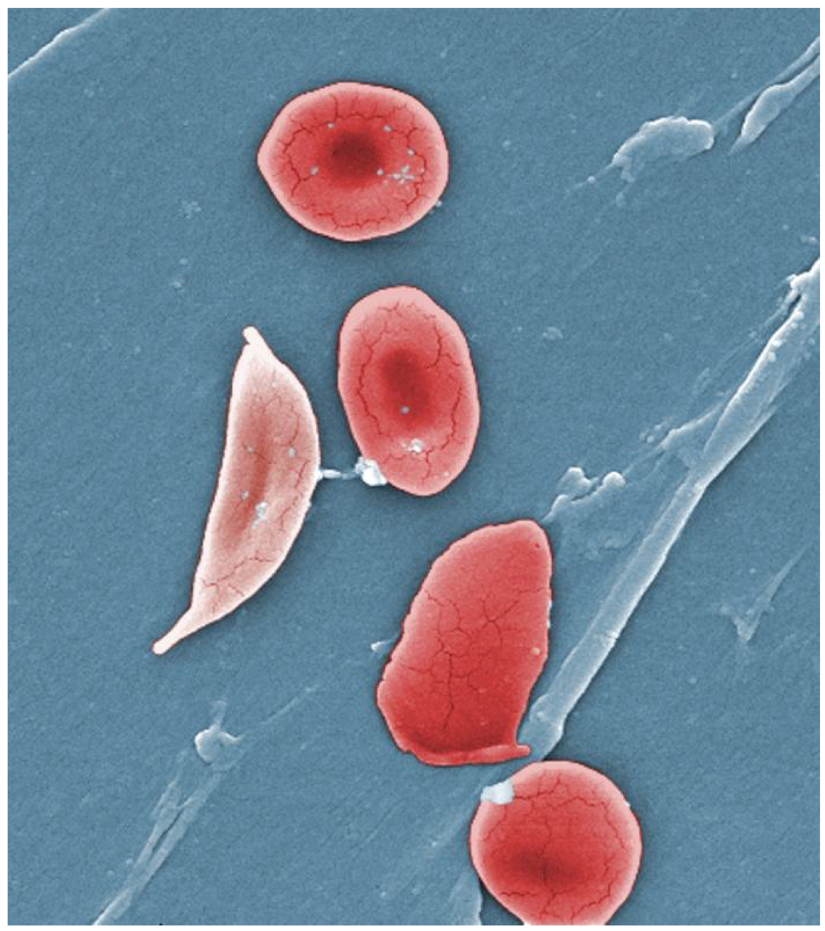
Additionally, they found that AOSLO imaging resolved single sickled red blood cells, rouleaux formations and blood cell-vessel wall interactions. These findings confirmed that the combined imaging modalities were sensitive enough to detail improved retinal perfusion in a sickle cell patient two months after initiation of oral hydroxyurea therapy.
“AOSLO imaging was able to reveal the cellular details of perfusion abnormalities detected using clinical OCT-A,” the study authors concluded. “The synergy between these clinical and laboratory imaging modalities presents a promising avenue in the management of sickle cell disease through development of noninvasive ocular biomarkers to prognosticate progression and measure response to systemic treatment.”
Pinhas A, Migacz JV, Zhou DB, et al. Insights into sickle cell disease through the retinal microvasculature: AOSLO correlates of clinical OCT angiography. Ophthalmology. July 11, 2022. [Epub ahead of print]. |