 |
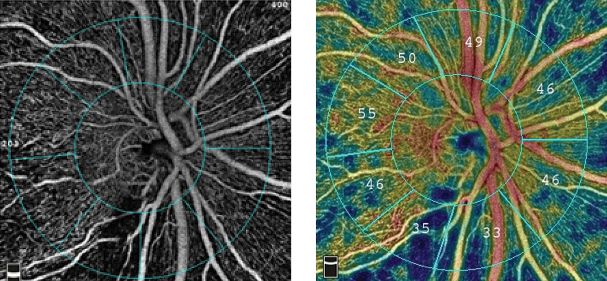
| OCT-A could provide you with useful information such as vessel density for monitoring PACG in affected patients. Photo: Julie Rodman, OD. Click image to enlarge. |
To help understand the vascular mechanism of glaucoma and improve diagnostic capability, researchers recently aimed to learn more about the clock-hour spatial characteristics of peripapillary vessel density using OCT angiography (OCT-A).
A total of 41 primary angle closure glaucoma (PACG) patients and 27 healthy participants took part in this cross-sectional study. Each underwent OCT-A and peripapillary retinal nerve fiber layer (RNFL) thickness imaging with swept-source OCT.
The study showed that peripapillary vessel density in the average, quadrant and clock-hour sectors of PACG decreased in different degrees compared with control eyes. The inferior quadrant vessel density had the highest diagnostic value. The diagnostic ability of vessel density in the four clock-hour and inferior quadrants were better than that of RNFL thickness.
“The reason may be that the severity of glaucoma in this study was much higher [than previous studies],” the authors explained in their paper. “Previous studies showed that peripapillary RNFL measured by OCT might not be as effective as visual field examination or macular thickness in monitoring the progression of advanced glaucoma.”
The authors noted an advantage of this study was the particular software they used to calculate the vessel density of each clock sector that matched the clock-hour RNFL sector. “We accurately matched clock-hour vessel density sectors and clock-hour RNFL sectors for the areas under the curve diagnostic capability comparison,” they noted.
Overall, the study suggested that OCT-A may provide useful information for glaucoma. “The diagnostic ability of vessel density in PACG was equivalent to that of traditional RNFL measurement,” the authors concluded in their study. “When the measurement of RNFL by OCT in advanced glaucoma becomes redundant, measuring microcirculation instead of nerve tissue itself may be more meaningful in monitoring the progress of diseases.”
Lin Y, Chen S, Zhang M. Peripapillary vessel density measurement of quadrant and clock-hour sectors in primary angle closure glaucoma using optical coherence tomography angiography. BMC Ophthalmol. September 9, 2021. [Epub ahead of print]. |