 |
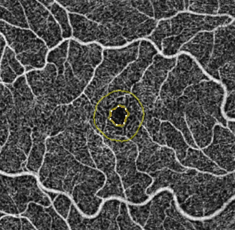
| FAZ circularity of the left eye was significantly lower in moderate, severe and PDR compared with no DR, whereas in right eyes these differences were only noticeable in severe and PDR compared with no DR. Photo: Julie Rodman, OD. Click image to enlarge. |
Retinal OCT-A metrics have provided additional markers for diabetic retinopathy (DR) assessment and allows precise investigation of microvascular changes in systemic diseases such as DR. Because of the differences of hemodynamic stress between right and left carotid arteries, researchers in Vienna hypothesized that this finding extends to retinal blood flow parameters in diabetic patients. Using OCT-A, they recently determined an independent inter-eye difference of vessel density and fractal dimension in the superficial capillary plexus (SCP), with the left eye showing reduced values compared with the right eye. Furthermore, foveal avascular zone (FAZ) circularity was more affected in left compared to right eyes.
A total of 336 eyes of 168 diabetic patients without DR and with DR stages ranging from mild nonproliferative to proliferative DR were included for analysis. Fovea-centered swept-source 6x6mm scans were acquired using a 200 kHz OCT-A device.
By analyzing absolute differences between eyes, the study found a significantly lower fractal dimension and vessel density in the SCP of the left compared to the right eye. This difference was independent of the DR stage and might be due to anatomic differences. FAZ circularity was lower in the left compared to right eyes without DR, moderate DR and proliferative DR (PDR). The researchers noted that this finding could indicate an earlier impairment of the FAZ microvasculature of the left eye.
Although left eyes were less circular in all, this difference failed to reach significance in patients with mild and severe DR. Higher FAZ perimeters were observed in the left compared the right eye in patients with PDR.
“By analyzing all eyes, we also found a reduction of the deep capillary plexus in advanced compared with early DR stages, which was not present in the SCP,” the researchers wrote in their paper. “However, the here-reported significant inter-eye difference in the SCP may be taken into account in future microvascular blood-flow analyses of the microvasculature of patients with DR.”
“Although we found a significant reduction of the SCP, as well as a more irregular FAZ in the left compared to the right eye, we can only speculate about the cause,” they added. “This inter-eye difference might be caused by higher hemodynamic stress to the left carotid artery and needs to be investigated in future studies.”
Stino H, Huber KL, Kunze L, et al. Intereye microvascular differences in patients with same-stage diabetic retinopathy revealed by OCT-A. Invest Ophthalmol Vis Sci. 2024;65(6):11. |