 |
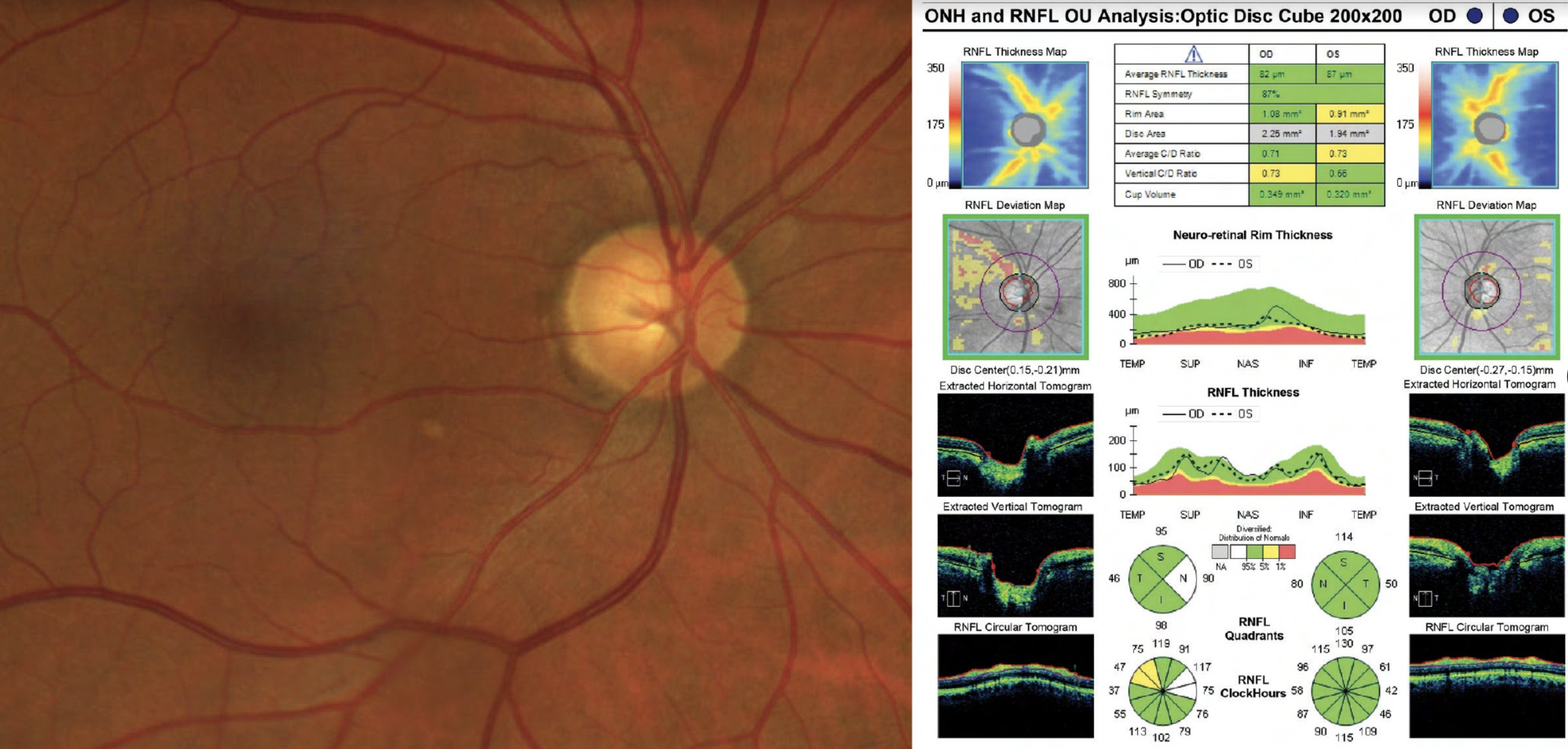
| Combined OCT and fundus imaging aid in glaucoma telemedicine screening. Photo: Ryan Schott, OD. |
The optimal protocols for glaucoma screening remain a point of debate, as some practitioners base their findings on multiple standard tests, while others have added imaging technology to their diagnostic toolbox. A new study that evaluated the accuracy of OCT imaging and fundus photography in a glaucoma telemedicine screening program found this combination resulted in a detection rate of about 9%, with moderate sensitivity, high specificity and predictive values that ranged from 84% to 96%.
The telemed screening program identified almost 20% of glaucoma suspects among individuals initially examined. The detection rate for confirmed glaucoma was about 5% and roughly 9% for confirmed or probable glaucoma.
The research team from Spain enrolled 4,113 participants from the Barcelona region who were scheduled for an exam at a primary care center. VA and IOP were measured. SD-OCT images of the optic disc, nerve fiber layer and ganglion cell complex at the macula were obtained, and fundus photography was used to image the optic disc and the macula. Exam results were uploaded to the telemedicine platform and sent for remote evaluation. All participants were then invited to attend a complete glaucoma exam, including gonioscopy, visual fields and dilated fundus evaluation.
In total, the investigators screened 1,006 individuals. Of these, 201 (about 20%) were classified as glaucoma suspects. Additionally, about 20% of participants were identified by retinography alone, roughly 13% by OCT imaging alone and approximately 46% by both.
Following the telemedicine screening, 481 individuals attended ophthalmic exams. During the in-person visit, 58 (12%) patients had confirmed glaucoma, 75 (16%) had probable glaucoma, 10 (2%) had ocular hypertension and 337 (70%) had no evidence of the condition.
The fundus photography and OCT imaging combo showed a sensitivity of 100% for advanced glaucoma cases and a sensitivity of 69% with a specificity of 95% for confirmed or probable cases, which could be the result of associating OCT with color retinography, the researchers suggested.
Additionally, during the screening process, 32% of participants had relevant findings in either photographs or OCT but not in both, suggesting the combination is more effective for screening purposes, the authors suggested.
“Overall, the results confirmed good diagnostic accuracy in a real-world screening environment and support the use of imaging devices for glaucoma screening,” they wrote in their paper.
Anton A, Nolivos K, Pazos M, et al. Diagnostic accuracy and detection rate of glaucoma screening with optic disk photos, optical coherence tomography images, and telemedicine. J Clin Med. 2021;11(1):216. |