 |
|
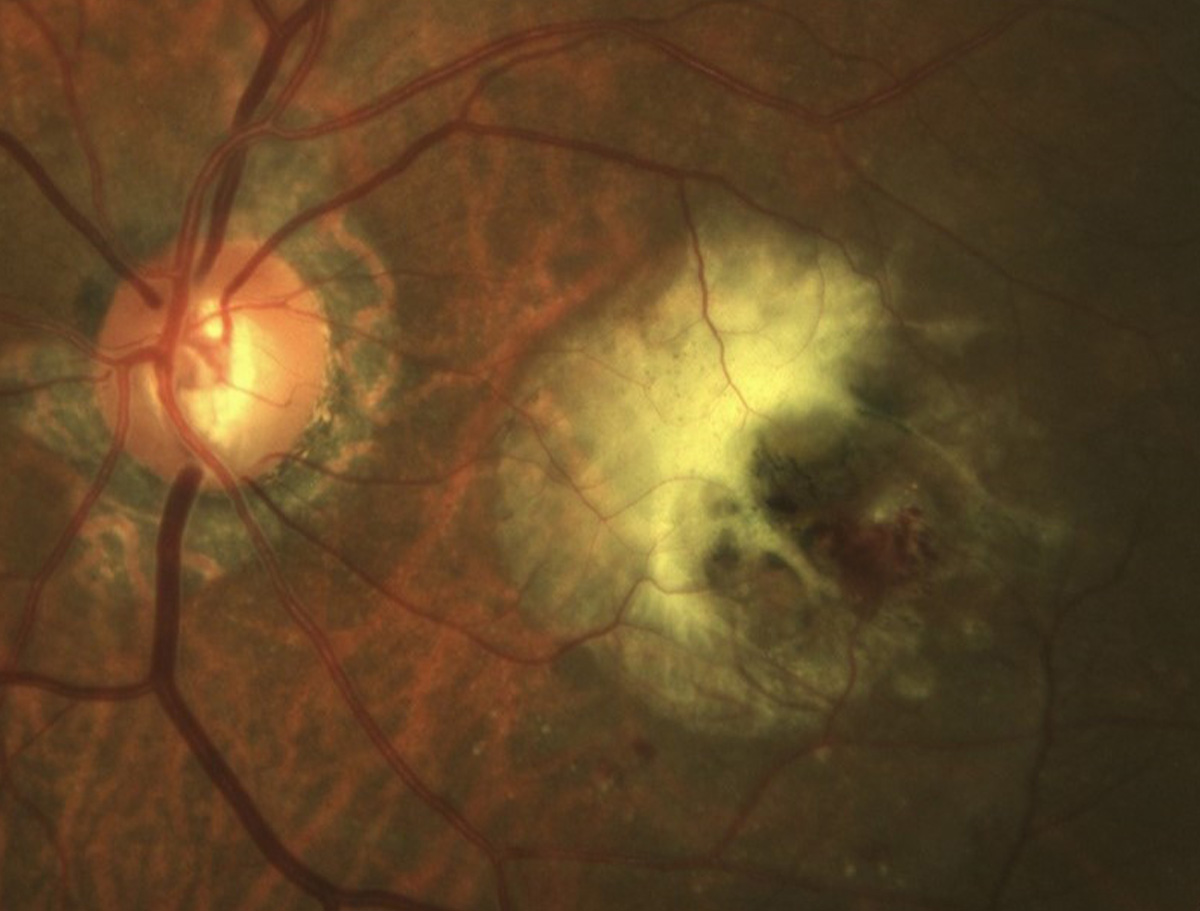
A recent analysis, which showed that fluid volume in the inner retina is an important predictor of short-term visual outcomes in Type 3 MNV, emphasizes the negative impact of intraretinal fluid on neuroretinal structures. Photo: Mohammad Rafieetary, OD. Click image to enlarge. |
Data from a retrospective cohort study demonstrated that the fluid volume in the inner retina is a key predictor of short-term visual outcomes among patients with Type 3 macular neovascularization (MNV).
“This study aims to quantify the volume of intraretinal fluid (IRF), subretinal fluid (SRF) and sub-retinal pigment epithelium (sub-RPE) fluid in treatment-naïve Type 3 MNV eyes with age-related macular degeneration (AMD) and to investigate the correlation of these fluid volumes with visual acuity outcomes at baseline and following anti-vascular endothelial growth factor (VEGF) treatment,” the study authors explained in their recently published American Journal of Ophthalmology paper.
In this analysis, the researchers analyzed 46 eyes from 46 patients with exudative AMD and treatment-naïve Type 3 MNV undergoing a loading dose of anti-VEGF therapy. A validated deep-learning segmentation strategy was used to process optical coherence tomography (OCT) B-scans, and segment and quantify IRF (i.e., both in the inner and outer retina), SRF and sub-RPE fluid volumes at baseline.
Among study participants, the investigators reported that visual acuity was 0.51±0.30 LogMAR at baseline and 0.33±0.20 LogMAR after the loading dose of anti-VEGF. At the follow-up visit, data showed a visual acuity of 0.40±0.17 LogMAR in patients with no complete resolution of retinal fluid and 0.31±0.20 LogMAR in those without retinal fluid after treatment.
Multivariable analysis revealed that the intraretinal fluid volume in the inner retina and the distance of the MNV from the fovea were predictors of visual acuity at baseline. The researchers found that baseline IRF volume in the inner retina also predicted the visual acuity at follow-up.
“In summary, the present study showed that eyes with Type 3 MNV are commonly complicated by IRF at baseline,” the study authors wrote in the American Journal of Ophthalmology. “Consequently, in such cases, quantifying the volume of fluid within retinal layers may hold prognostic significance, as our results suggest a potential correlation between the amount of fluid in the inner retina and visual outcomes.
“This observation aligns with speculation in other disorders where inner retinal fluid presence may disrupt connections between photoreceptors and ganglion cells, possibly leading to visual impairment,” they concluded. “Future prospective studies with extended follow-up periods could validate our findings and ascertain whether the quantity of retinal fluid in the inner retina might serve as a biomarker for long-term visual outcomes, complementing its role in short-term visual prognostication.”
Berni A, Oakley JD, Dolz-Marco R, et al. Topographical Quantification of Retinal Fluid in Type 3 MNV and Associations with Short-Term Visual Outcomes. Am J Ophthalmol. August 30, 2024 [Epub ahead of print]. |