 |
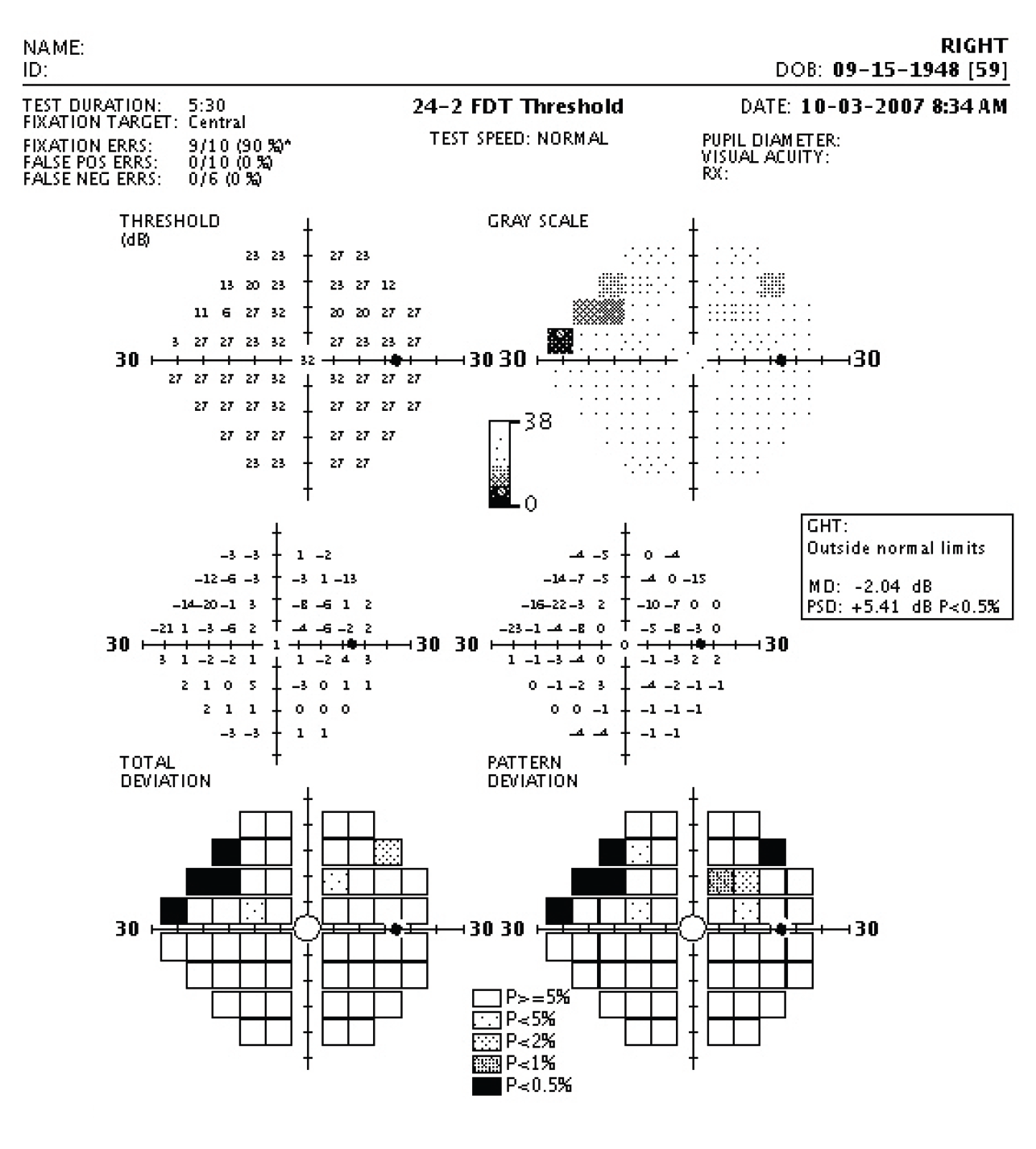
| This study found that a greater difference in frequency-doubling technology perimetry and standard automated perimetry was associated with structural parameters. Photo: Carl Zeiss Meditec. Click image to enlarge. |
Standard automated perimetry (SAP) is the most common method for detecting visual field damage in glaucoma patients, but this method is not as effective in detecting early changes, which are often more structural than functional. One proposed alternative is frequency-doubling technology (FDT) perimetry, which is based on the frequency-doubling illusion where each stimulus is a series of white and black lines that flicker at 25Hz, as opposed to the SAP white stimulus on a white background.
“FDT perimetry is thought to be mediated by a subset of retinal ganglion cells with large axonal diameter, called the M6y ganglion cells, that project to the magnocellular pathway. These cells are sensitive to motion and contrast and are thought to be more vulnerable to mechanical glaucomatous damage than conventional perimetry,” explained researchers of a recent study.
They investigated the ability of FDT and SAP to detect visual field damage, finding that the difference between these two methods may be useful in identifying risk factors in glaucoma patients.
Their cross-sectional study included early-stage glaucoma patients (n=96 eyes with mean deviation >-6dB). Mean deviation and pattern standard deviation of FDT were subtracted from those of SAP, and several clinical features were measured. Here are some of the major findings:
Eyes with significant SAP-FDT difference demonstrated higher detection of microvasculature dropout on the deep-layer OCT-A map, greater lamina cribrosa depth and greater lamina cribrosa curvature index.
Logistic regression analysis found that frequent presence of microvascular dropout, less presence of disc hemorrhage and greater lamina cribrosa depth were significantly associated with SAP-FDT difference.
In a sub-analysis of peripheral nasal step eyes (n=50) and parafoveal scotoma eyes (n=46), the SAP-FDT difference of the mean deviation value demonstrated a positive association with peripapillary vessel density on deep-layer OCT-A. This was significant in parafoveal scotoma eyes vs. peripheral nasal step eyes.
The SAP-FDT difference in pattern standard deviation value had a negative association with lamina cribrosa curvature index and lamina cribrosa depth. This was significant in peripheral nasal step eyes vs. parafoveal scotoma eyes.
To summarize, the researchers wrote that a greater difference in SAP and FDT was associated with structural parameters, such as lamina cribrosa depth and lamina cribrosa curvature index. Less of a difference was associated with the presence of disc hemorrhage and lower deep-layer peripapillary vessel density.
“In clinical practice, it may help to predict associated risks and make plans for glaucoma evaluation and treatment by comparing FDT and SAP,” they wrote.
Kim S, Park C, Park HYL, et al. Comparison between frequency-doubling technology perimetry and standard automated perimetry in early glaucoma. Sci Rep. 2022;12(1):10173. |