Swedish researchers recently investigated the characteristics of optic nerve hypoplasia (ONH) in a small group of patients with the congenital condition of different severities. Using high-resolution spectral domain OCT, a total of 36 patients (52 ONH eyes and 17 fellow eyes in unilateral cases) as well as 45 healthy right eyes from 45 controls were evaluated.
After analyzing images of the disc and macula, researchers were able to determine that ONH eyes upon OCT had a shorter disc diameter (1,061µm ±375µm vs. 1,751µm ±221µm), shallower mean cup depth (427µm ±171µm vs. 551µm ±152µm), thinner ganglion cell complex (GCC) perifoveally (47.3µm ±13.0µm vs. 60.8µm ±6.0µm) and reduced foveal depth (61µm ±36µm vs. 119µm ±19µm) when compared with control eyes.
 |
|
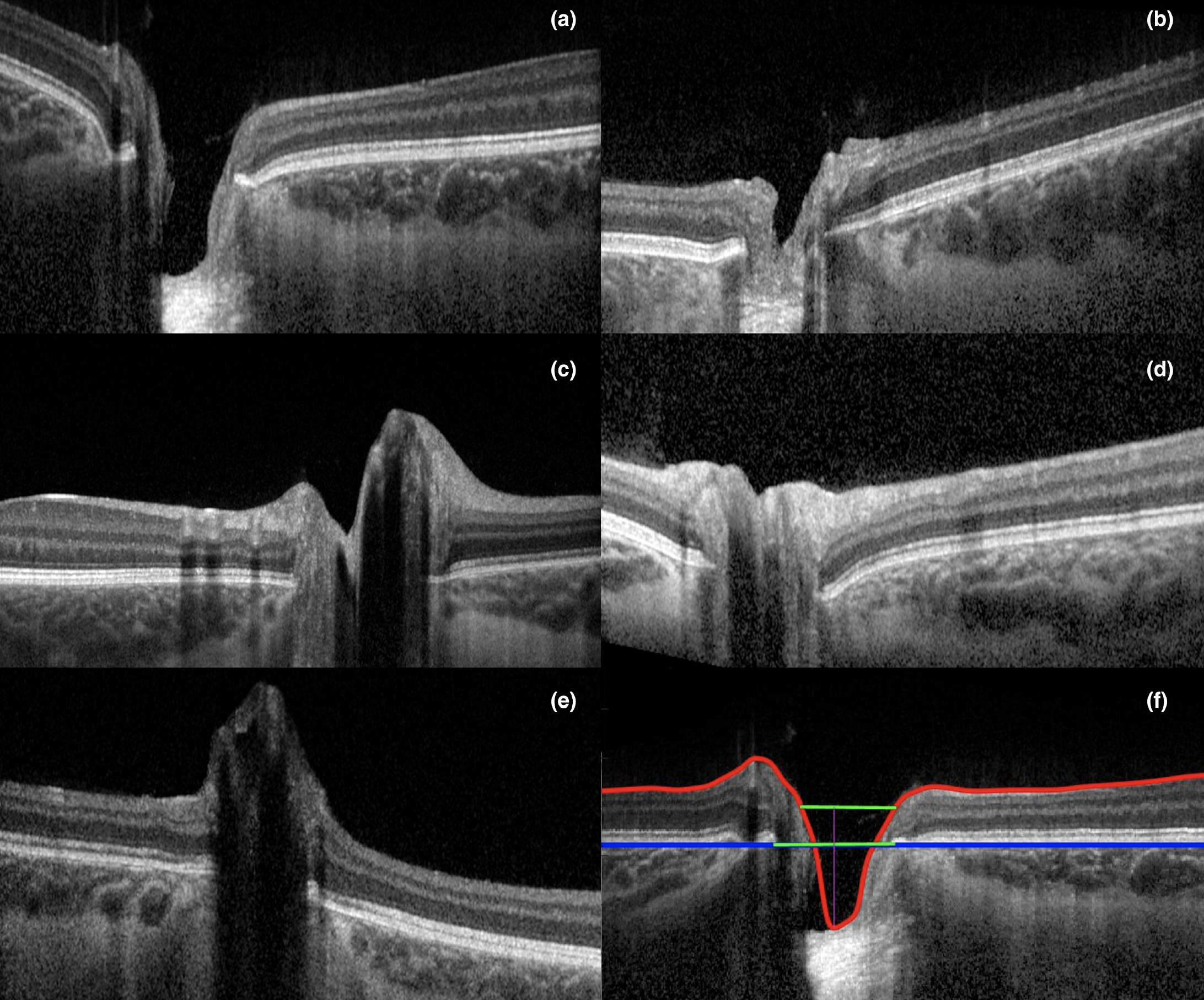
The study group saw about 30% of eyes with a ‘subnormal’ fovea and lower prevalence of foveal hypoplasia (35%) than another study. These images of study participants show a wide range of physiological feature of optic nerve hypoplasia:(a) cup below RPE, (b) cup below IRL, (c) cup above IRL, (d) no cup and (e) peak formation. Image (f) shows demarcations of the ILM, RNFL, RPE and Bruch’s membrane opening. Photo: Skriapa-Manta A, et al. Acta Ophthalmol. May 23, 2024. Click image to enlarge. |
Upon qualitative analysis, one-third of ONH eyes lacked signs of an optic cup while two-thirds displayed reduced or no sign of a foveal pit. Interestingly, fellow eyes of those with ONH had shorter disc diameter (1,446µm ±404µm vs. 1,751µm ±221µm) and reduced foveal depth (93µm ±27µm vs. 119µm ±19µm); however, similar GCC thickness was observed (60.8µm ±7.1µm vs. 60.8µm ±6.0µm) compared with controls. Visual acuity in ONH eyes was best correlated with disc diameter.
Along with these two new presentations of ONH eyes, the smaller disc diameter and reduced foveal depth of fellow eyes in presumed unilateral cases suggests possible subclinical or mild disease. This, along with the normal GCC thickness of these cases, prompted the authors to stress that “the correlation between structure and visual function is not always straightforward.”
In their discussion, the authors mention how a large part of difference seen in mean cup depth was due to a third of ONH eyes lacking measurable optic cup depth—a rare occurrence in fellow eyes and nonexistent in control eyes—but this may not be true of the entire population. Consequently, lack of measurable optic cup was highly predictive of severe ONH.
The observations seen in fellow eyes of shallower foveal depth and reduced disc diameter are features that could easily be missed upon ordinary clinical examination without performance of OCT. As such, the authors recommend that “OCT evaluations should be part of a standard clinical protocol in ONH evaluation. The fellow eyes could have subclinical disease, and bilateral ONH has been shown to be more frequently associated with comatic, hormonal or extraocular manifestations.”
They then contextualize the larger theme of their results, stating that “the larger overlap in various OCT parameters between ONH eyes, fellow eyes and healthy eyes due to the expanded spectrum of disease severity highlights the complexity of the disorder.”
“OCT is a useful tool in diagnosing most patients with ONH,” they add. “However, equivocal cases still exist and need a thorough evaluation, including multimodal imaging, to confirm the diagnosis.”
Skriapa-Manta A, Venkataraman AP, Olsson M, Nilsson M, Fahnehjelm KT. Characteristic deviations of the optic disc and macula in optic nerve hypoplasia based on OCT. Acta Ophthalmol. May 23, 2024. [Epub ahead of print]. |